Translate this page into:
Telltale signs of skin trespassers: Clues to superficial mycosis
Corresponding author: Dr. Varsha Manche Gowda, Department of Dermatology, Venereology and Leprosy, Bangalore Medical College and Research Institute, Bengaluru, Karnataka, India. varshamgowda95@gmail.com
-
Received: ,
Accepted: ,
How to cite this article: Varsha MG, Shilpa K, Revathi TN, Shanmukhappa AG, Loganathan E. Telltale signs of skin trespassers: Clues to superficial mycosis. Indian J Dermatol Venereol Leprol 2023;89:144-8.
Superficial fungal infections constitute the commonest reason for a dermatologic consultation across India. However, recently they have become a diagnostic challenge due to various reasons. This article highlights certain signs and appearances that may facilitate their diagnosis.
Clinical Signs
Besnier’s sign/scratch sign/stroke of the nail/coup d’ongle sign
The fine branny scales of pityriasis versicolor may become visible after scratching the lesions with a sharp object. This sign is negative in treated cases and following a recent bath [Figure 1].1

- Besnier’s sign in pityriasis versicolor. (a) Before scratching. (b) After scratching with fingernail
Double-edged Tinea/Ring-within-Ring Appearance
It is an important clinical marker of steroid modified tinea.2 Due to relapsing inflammation, there is incomplete clearance of the fungal elements which clinically presents as parallelly arranged erythematous borders. This creates a double-edged appearance and the annular lesions develop multiple concentric rings spreading centrifugally, appearing like ‘ring within ring’ [Figure 2].3

- Steroid modified tinea. (a) Double-edged tinea. (b) ‘Ring within ring’ appearance
Dumbbell-shaped Tinea
This appearance also indicates tinea incognito, characterised by the confluence of multiple annular lesions of various sizes across multiple anatomical locations. Additionally, there is loss of central clearing with eczematisation, creating a ‘dumbbell’ appearance [Figure 3].2,3

- (a) Dumbbell-shaped tinea. (b) Both dumbbell and ring within ring appearance seen
Ear Sign
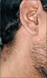
Ear sign denotes the presence of erythematous scaly papules and plaques over helix, antihelix and retroauricular region, sparing the retroauricular fold, classical of tinea capitis. Notably, identical lesions involving the retroauricular folds indicate seborrheic dermatitis.4 Recently, ‘ear sign’ has also been attributed to the involvement of auricular pinna, suggestive of ipsilateral tinea faciei [Figure 4].3

- Ear sign. Auricular pinna involved in a case of tinea faciei
Egg Crackling Sign
This clinical sign has been described in elderly patients with pityriasis versicolor. On stretching the lesion, the overlying scales break like the crackling of an eggshell.
Salt and Pepper Appearance
Tinea nigra is a superficial fungal infection characterised by asymptomatic brown to black macules and patches over palms and soles. A rare variant has been described which forms the speckled or ‘salt and pepper’ pattern over the palms.5
Zireli’s Sign/Evoked Scale Sign/Stretch Sign
On stretching the lesions of pityriasis versicolor with two fingers at 180° angle, the corneal layer parasitised by Malassezia gets exposed.6 This sign helps to differentiate pityriasis versicolor and other disorders involving dyspigmentation [Figure 5].7

- Zireli’s sign in pityriasis versicolor. (a) Before, (b) during and (c) after stretching showing evident scales
Dermoscopic Signs
Aurora borealis pattern
On onychoscopy, onycholytic nail plate demonstrates multiple longitudinal lines of varied colours (yellow, brown, white, etc.) similar to the colours of aurora borealis. They represent fungal colonies, subungual debris and invasion, and is most commonly observed in distal and lateral subungual onychomycosis [Figure 6].8

- Aurora borealis sign of onychomycosis showing multicoloured pigmentation on onychoscopy (×10, DL4, polarised view)
Cigarette-ash Hair
This sign indicates post-treatment cases of tinea capitis. As the endothrix spores are eliminated by antifungals, the corkscrew hair become more fragile and easily breakable. New uninfected hair replaces them, which resemble cigarette ash on dermoscopy.9
Comma Hairs
It is a trichoscopic feature of tinea capitis. Multiple hyphae fill the hair follicle resulting in cracking and bending of the hair shaft with a marked distal angulation resembling ‘comma’ [Figure 7].10

- Trichoscopy of tinea capitis showing comma hair (white circles), corkscrew hair (blue circles), telephone handle hair (red circles) and zigzag hair (arrow) (×10)
Contrast Halo Sign
Dermoscopy of pityriasis versicolor reveals increased pigmentation around hypopigmented lesions and identical decreased pigmentation around primary lesions of hyperpigmented variant. This is called ‘Contrast halo sign’ [Figure 8].

- Contrast halo sign. A rim of hyperpigmentation (black arrows) around a hypopigmented lesion of pityriasis versicolor (×10, DL4, polarised view)
Proposed mechanism – In hypopigmented variant, the compensatory melanogenesis occurs due to cytotoxic damage and abnormal melanosomes in the primary lesion, while in hyperpigmented variant, the consumption of melanocytes occurs as a response to the stimulated melanogenesis in the primary lesion due to perivascular inflammation.11
Corkscrew Hair
Corkscrew hair is considered to be a trichoscopic indicator of tinea capitis. Fungal invasion of a hair follicle along with its continuous growth results in bending and coiling of hair [Figure 7].9
Fish Net/Wire Fence Appearance
It is an easy and quick dermoscopic clue to pityriasis versicolor, useful when scratch sign is clinically negative. It is characterised by fine scales along the normal skin markings against a hypo/hyperpigmented background, simulating a ‘wire fence’/‘fish net’. [Figure 9].12

- Fishnet/wire fence appearance, folliculocentricity of scales seen on dermoscopy of pityriasis versicolor (×10, DL4, non-polarised view)
Morse Code/Bar Code Hair
It is a recent dermoscopic feature reported in tinea capitis and corporis, appearing as subtle interrupted horizontal white bands due to localised fungal invasion.13 These bands are multiple and represent ‘locus minoris resistentiae’ that make the hair easily deformable, translucent and fragile [Figure 10].14

- Morse code hair seen on dermoscopy of tinea corporis as horizontal light bands resulting in bending of hair (arrows) (×10, DL4, polarised view)
Motheaten Scales
It is a specific dermoscopic feature of tinea corporis. Motheaten scales represent multiple annular lesions with peripheral scaling, coalescing to form larger multicyclic lesion in outward peeling direction [Figure 11].15

- Dermoscopy of tinea corporis showing peripheral interrupted motheaten scales with background erythema (×10, DL4, non-polarised view)
Reverse Triangular Pattern
This pattern is described in onychoscopy of onychomycosis. The black reverse triangle forms as wider nail pigmentation occurs at the distal end indicating fungal invasion from that end. The triangular sign is also observed in ungual melanoma and nail matrix nevus in children [Figure 12].16

- Onychoscopy of onychomycosis showing reverse triangular sign (red triangle) (×10, DL4, polarised view)
Ruin Appearance
Ruin appearance is the special dermoscopic term depicting subungual keratosis in onychomycosis. It denotes the indentations of the ventral nail plate which occur due to accumulation of dermal debris in response to fungal invasion. It is classical for total dystrophic onychomycosis [Figure 13].17

- Onychoscopy of onychomycosis showing ruin appearance (×10, DL4, non-polarised view)
Telephone Handle Hair
It is a novel trichoscopic finding in tinea capitis. Due to fungal invasion, the hair shafts become easily deformable, horizontally bent with slightly bulbous appearance on either side resembling a ‘telephone handle’ [Figure 7].18
Zigzag Hair
It is another common trichoscopic feature in tinea capitis. The conidia on hair surface following fungal invasion of hair cuticle bends the paler part of infected hair resulting in its structural weakness and zigzag hair [Figure 7].10
Laboratory Signs
Butter cream frosting appearance
It describes the colony appearance of Trichosporon spp., the causative organism of white piedra. On Sabouraud’s dextrose agar, rapid growth of moist cream-coloured yeast-like colonies resemble ‘butter cream frosting.’19
Mosaic Fungus
It is the most common artefact encountered during direct microscopic examination of skin scrapings for fungal elements. KOH dissolves normal epidermal cells forming irregular branching network resembling fungal structures [Figure 14].20

- KOH image showing mosaic fungus (×40)
Sandwich Sign
Histopathological examination of dermatophytosis demonstrates fungal elements ‘sandwiched’ between the two zones of stratum corneum, upper orthokeratotic and lower parakeratotic layer [Figure 15].1

- Sandwich sign. Histopathology with PAS staining of tinea corporis showing fungal hyphae in the stratum corneum (black arrows) (×40)
Spaghetti and Meatballs/Banana and Grapes Appearance
On KOH mount examination of scales of pityriasis versicolor, thick-walled spherical yeasts of Malassezia furfur are present in clusters with scattered short septate filaments (2–5 μ wide, 25 μ long) resembling ‘banana and grapes’ or ‘spaghetti and meatballs’. [Figure 16].20

- KOH image of pityriasis versicolor showing hyphae as spaghetti (white arrows) and spores as meatballs (black arrows) (×40)
Declaration of patient consent
The authors certify that they have obtained all appropriate patient consent.
Financial support and sponsorship
Nil.
Conflicts of interest
There are no conflicts of interest.
References
- Eponymous signs in dermatology. Indian Dermatol Online J. 2012;3:159-65.
- [CrossRef] [PubMed] [Google Scholar]
- The great Indian epidemic of superficial dermatophytosis: An appraisal. Indian J Dermatol. 2017;62:227-36.
- [Google Scholar]
- The unprecedented epidemic-like scenario of dermatophytosis in India: I. Epidemiology, risk factors and clinical features. Indian J Dermatol Venereol Leprol. 2021;87:154-75.
- [CrossRef] [PubMed] [Google Scholar]
- Useful sign to diagnose tinea capitis-“ear sign”. Indian J Pediatr. 2012;79:679-80.
- [CrossRef] [PubMed] [Google Scholar]
- Tinea nigra presenting speckled or “salt and pepper” pattern. Am J Trop Med Hyg. 2014;90:981.
- [CrossRef] [PubMed] [Google Scholar]
- White piedra, black piedra, tinea versicolor, and tinea nigra: Contribution to the diagnosis of superficial mycosis. An Bras Dermatol. 2017;92:413-6.
- [CrossRef] [PubMed] [Google Scholar]
- Diagnosis of pityriasis versicolor in paediatrics: The evoked scalesign. Arch Dis Child. 2011;96:392-3.
- [CrossRef] [PubMed] [Google Scholar]
- Diagnostic utility of onychoscopy: Review of literature. Indian J Dermatopathol Diagn Dermatol. 2017;4:31-40.
- [CrossRef] [Google Scholar]
- An ultrastructural study on corkscrew hairs and cigarette-ash-shaped hairs observed by dermoscopy of tinea capitis. Scanning. 2016;38:128-32.
- [CrossRef] [PubMed] [Google Scholar]
- Tinea capitis in children: A report of four cases trichoscopic with trichoscopic features. Indian J Paediatr Dermatol. 2018;19:51-6.
- [CrossRef] [Google Scholar]
- Dermoscopy in the evaluation of pityriasis versicolor: A cross sectional study. Indian Dermatol Online J. 2019;10:682-5.
- [CrossRef] [PubMed] [Google Scholar]
- Dermoscopy: An easy way to solve the diagnostic puzzle in pityriasis versicolor. Indian J Dermatol Venereol Leprol. 2019;85:664-5.
- [CrossRef] [PubMed] [Google Scholar]
- Idiosyncratic findings in trichoscopy of tinea capitis: Comma, zigzag hairs, corkscrew, and Morse code-like hair. Int J Trichol. 2016;8:180-3.
- [CrossRef] [PubMed] [Google Scholar]
- Can dermoscopy serve as a diagnostic tool in dermatophytosis? A pilot study. Indian Dermatol Online J. 2019;10:530-5.
- [CrossRef] [PubMed] [Google Scholar]
- Dermatoscopy of tinea corporis. J Eur Acad Dermatol Venereol. 2020;34:e278-80.
- [CrossRef] [PubMed] [Google Scholar]
- Dermoscopic patterns of fungal melanonychia: A comparative study with other causes of melanonychia. J Am Acad Dermatol. 2017;76:488-93.
- [CrossRef] [PubMed] [Google Scholar]
- Dermoscopy as a first step in the diagnosis of onychomycosis. Postepy Dermatol Alergol. 2018;35:251-8.
- [CrossRef] [PubMed] [Google Scholar]
- Telephone handle hair: A novel trichoscopic finding in black dot tinea capitis. Int J Trichol. 2019;11:181-3.
- [CrossRef] [PubMed] [Google Scholar]
- Fungal diseases In: Bolognia JL, Schaffer JV, Cerroni L, eds. Dermatology Vol 2. (4th ed). Amsterdam, Netherlands: Elsevier; 2012. p. :1330-1.
- [Google Scholar]
- Skin scraping and a potassium hydroxide mount. Indian J Dermatol Venereol Leprol. 2006;72:238-41.
- [CrossRef] [PubMed] [Google Scholar]


