Translate this page into:
Treatment of congenital melanocytic nevi in the periorbital area with dual-wavelength copper vapor laser
Corresponding author: Dr. Igor V. Ponomarev, Head of Laser Project, 53, Leninskiy Prospect, Moscow, 119991, Russian Federation. luklalukla@ya.ru
-
Received: ,
Accepted: ,
How to cite this article: Ponomarev IV, Topchiy SB, Andrusenko YN, Shakina LD. Treatment of congenital melanocytic nevi in the periorbital area with dual-wavelength copper vapor laser. Indian J Dermatol Venereol Leprol 2021;87:720-2.
Sir,
Facial congenital melanocytic nevus represents a rare condition with a prevalence of about 2%.1 As periorbital congenital melanocytic nevus appears as a hyperpigmented plaque close to the eye, it inevitably poses cosmetic problems. The excision of the nevus is reported to be associated with the high risk of scarring in the esthetic zones.2 Ablative lasers such as CO2 laser (10,600 nm) and Er:YAG (2940 nm) as well as non-ablative mid-infrared lasers, especially ruby (694 nm), alexandrite (755 nm) and Nd:YAG (1064 nm) have so far been successfully tried for the treatment of congenital melanocytic nevus.3,4 Nevertheless, the application of laser systems in the periorbital area remains limited due to reported side effects including bleeding, edema, relapses, scarring and ocular complications.4 The complications of laser treatment may emerge due to the deep penetration of mid-infrared radiation because of its low absorption by both melanin and hemoglobin assumed as targeted chromophores. However, dual-wavelength copper vapor laser radiation at a wavelength of 511 nm is highly absorbed by melanin and radiation at 578 nm is mostly absorbed by hemoglobin. Thus, this laser seems to be the most appropriate for the safe removal of periorbital congenital melanocytic nevus.
The nevi were treated using copper vapor laser in two patients with Fitzpatrick II skin phototype. A 14-year-old girl presented with a brownish plaque of size 42 × 35 mm beneath the left lower eyelid [Figure 1]. A 23-year-old man presented with, a hyperpigmented plaque of size 30 × 50 mm in the right periorbital area [Figure 2]. The nevi were noted since birth and increased in size during body growth. No history of melanoma was present in patients or their relatives. Based on clinical signs (size [M], location, the number of satellites [S], color uniformity [C], roughness [R], nodules [N] and hair [H]) and dermoscopic data, the diagnosis of congenital melanocytic nevus Type I, M1S0C1R1N0H0 was made.
In both cases, the diagnosis was confirmed by skin biopsy which showed accumulated mature nevomelanocytes in the superficial and mid-dermis. Both patients gave informed consent for the treatment of the nevi using laser.
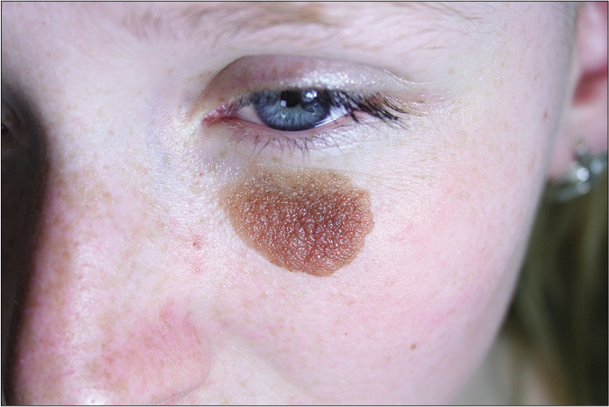
- The 14-year-old girl with congenital melanocytic nevus at the left periorbital area before copper vapor laser treatment
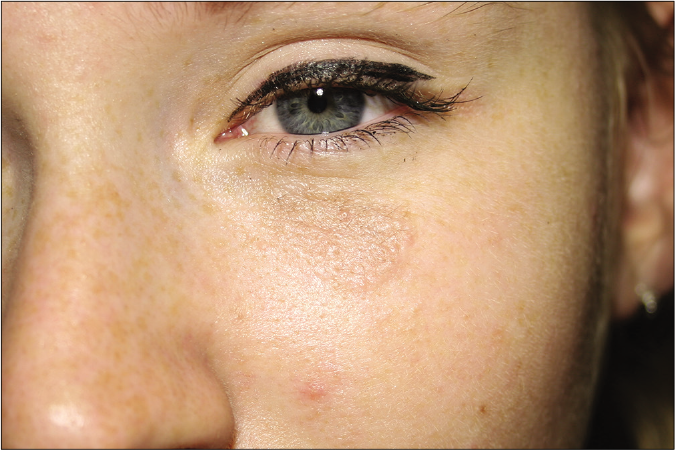
- The 14-year-old girl with congenital melanocytic nevus at the left periorbital area after the third copper vapor laser treatment
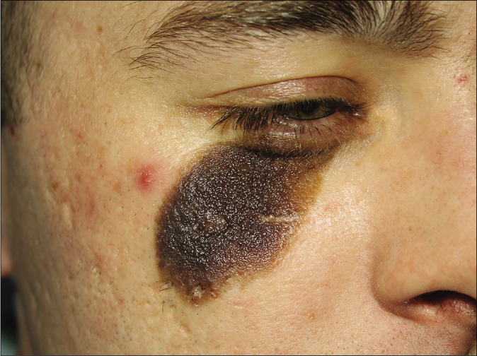
- The 23-year-old man with congenital melanocytic nevus on the right periorbital area before the copper vapor laser treatment
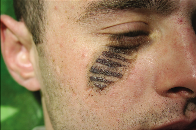
- The 23-year-old man with congenital melanocytic nevus 1 month after the first copper vapor laser treatment
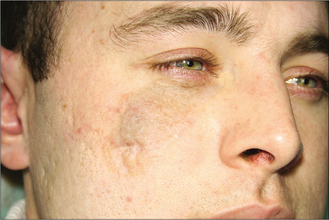
- The 23-year-old man with congenital melanocytic nevus 4 months after the third copper vapor laser treatment
Dual-wavelength radiation of the copper vapor laser settings was chosen as follows: power ratio at wavelengths of 511 nm and 578 nm was selected in a 3:2 mode, the average power – 0.7 W and 0.8 W for the first and second cases respectively, exposure time – 0.3 s and light spot diameter at the skin – 1 mm. The treatment was performed using a multiple stacking pass technique. The treatment endpoint was set when the irradiated area of the nevi acquired a grayish tint. The area was divided into equal transverse strips of 3–4 mm in width to provide sound healing [Figure 2b]. At first, just the odd strips were irradiated. One month later, after the healing of the already irradiated area, the even strips were irradiated. Both patients received three sessions with a one-month interval.
Laser treatment was performed without anesthesia. The immediate endpoint was accepted as the mild graying of the exposed area. No bleeding or erythema was noted during and after the laser procedure. After the laser procedure, the exposed skin was treated with 0.05% solution of chlorhexidine gluconate. In the early post-operative period, Bepanthen cream was applied twice a day. The skin of the irradiated area remained grayish for several days. In 7–10 days, there was exfoliation of crusts followed by the regeneration of epidermis without leaving behind any hyperpigmentation. Two weeks after the laser procedure, the irradiated area became entirely similar to adjoining intact skin [Figures 1b and 2c]. The healing process took 12 days. Patients were advised to use a broad-spectrum sunscreen during the treatment period and for a month after the last procedure. Both cases did not show any side effects during a 36-month follow-up period.
Ablative lasers (CO2 and Er:YAG) showed promise in treating periorbital congenitial melanocytic nevus, but side effects such as bleeding, scarring and recurrences were reported.3 The introduction of the above mentioned Q-switched pigment-specific mid-infrared laser into the clinical practice has made it possible to treat congenital melanocytic nevus with a lower complication rate, but there is a risk of persistent erythema, edema, relapse and risk of eye injury.4 A combination of intense pulsed light with Er:YAG laser has not proved to be a reliable treatment for this condition because of high recurrence.5
Our data prove that the copper vapor laser seems to be the optimal approach to the treatment of periorbital congenital melanocytic nevus due to the high absorption of the 511 nm radiation by melanin. Melanin absorbs the radiation with a wavelength of 511 nm, 10 times more than the radiation with 1064 nm wavelength. Hence, selective photodestruction of melanosome with copper vapor laser requires less light energy than the mid-infrared laser. The high absorption of 578 nm radiation by oxyhemoglobin and hemoglobin as well as high absorption of 511 nm radiation by melanin limits the penetration depth of the laser and protects the underlying eye structures from injuries.6
Remodeling of the vascular bed through selective vessel coagulation under the radiation with a wavelength of 578 nm prevents post-irradiation bleeding and relapse of congenital melanocytic nevus.
In our case series split-face comparative assessment of different laser treatment modes was not done and this can be factored as a limitation. More studies are required to determine the optimal green/yellow ratio of the copper vapor laser power for different clinical variants of congenital melanocytic nevus.
Declaration of patient consent
The authors certify that they have obtained all appropriate patient consent.
Financial support and sponsorship
Nil.
Conflicts of interest
There are no conflicts of interest.
References
- Clinico epidemiological study of cutaneous manifestations in the neonate. Indian J Dermatol Venereol Leprol. 2000;66:26-8.
- [Google Scholar]
- Serial excision with power stretching for large and giant melanocytic nevi of the trunk. J Dtsch Dermatol Ges. 2019;17:852-5.
- [CrossRef] [Google Scholar]
- Treatment of congenital melanocytic nevi in the eyelid and periorbital region with ablative lasers. Ann Plast Surg. 2019;83:565-9.
- [CrossRef] [Google Scholar]
- Laser treatment of congenital melanocytic nevi: A review of the literature. Lasers Med Sci. 2016;31:197-204.
- [CrossRef] [Google Scholar]
- Intense pulsed light alone and in combination with erbium yttrium-aluminum-garnet laser on small-to-medium sized congenital melanocytic nevi: Single center experience based on retrospective chart review. Ann Dermatol. 2017;29:39-47.
- [CrossRef] [Google Scholar]
- Numerical modeling and clinical evaluation of pulsed dye laser and copper vapor laser in skin vascular lesions treatment. J Lasers Med Sci. 2019;10:44-9.
- [CrossRef] [Google Scholar]


