Generic selectors
Exact matches only
Search in title
Search in content
Post Type Selectors
Search in posts
Search in pages
Filter by Categories
15th National Conference of the IAOMFP, Chennai, 2006
Abstract
Abstracts from current literature
Acne in India: Guidelines for management - IAA Consensus Document
Addendum
Announcement
Art & Psychiatry
Article
Articles
Association Activities
Association Notes
Award Article
Book Review
Brief Report
Case Analysis
Case Letter
Case Letters
Case Notes
Case Report
Case Reports
Clinical and Laboratory Investigations
Clinical Article
Clinical Studies
Clinical Study
Commentary
Conference Oration
Conference Summary
Continuing Medical Education
Correspondence
Corrigendum
Cosmetic Dermatology
Cosmetology
Current Best Evidence
Current Issue
Current View
Derma Quest
Dermato Surgery
Dermatopathology
Dermatosurgery Specials
Dispensing Pearl
Do you know?
Drug Dialogues
e-IJDVL
Editor Speaks
Editorial
Editorial Remarks
Editorial Report
Editorial Report - 2007
Editorial report for 2004-2005
Errata
Erratum
Focus
Fourth All India Conference Programme
From Our Book Shelf
From the Desk of Chief Editor
General
Get Set for Net
Get set for the net
Guest Article
Guest Editorial
History
How I Manage?
IADVL Announcement
IADVL Announcements
IJDVL Awards
IJDVL AWARDS 2015
IJDVL Awards 2018
IJDVL Awards 2019
IJDVL Awards 2020
IJDVL International Awards 2018
Images in Clinical Practice
Images in Dermatology
In Memorium
Inaugural Address
Index
Knowledge From World Contemporaries
Leprosy Section
Letter in Response to Previous Publication
Letter to Editor
Letter to the Editor
Letter to the Editor - Case Letter
Letter to the Editor - Letter in Response to Published Article
LETTER TO THE EDITOR - LETTERS IN RESPONSE TO PUBLISHED ARTICLES
Letter to the Editor - Observation Letter
Letter to the Editor - Study Letter
Letter to the Editor - Therapy Letter
Letter to the Editor: Articles in Response to Previously Published Articles
Letters in Response to Previous Publication
Letters to the Editor
Letters to the Editor - Letter in Response to Previously Published Articles
Letters to the Editor: Case Letters
Letters to the Editor: Letters in Response to Previously Published Articles
Media and news
Medicolegal Window
Messages
Miscellaneous Letter
Musings
Net Case
Net case report
Net Image
Net Images
Net Letter
Net Quiz
Net Study
New Preparations
News
News & Views
Obituary
Observation Letter
Observation Letters
Oration
Original Article
ORIGINAL CONTRIBUTION
Original Contributions
Pattern of Skin Diseases
Pearls
Pediatric Dermatology
Pediatric Rounds
Perspective
Presedential Address
Presidential Address
Presidents Remarks
Quiz
Recommendations
Regret
Report
Report of chief editor
Report of Hon : Treasurer IADVL
Report of Hon. General Secretary IADVL
Research Methdology
Research Methodology
Resident page
Resident's Page
Resident’s Page
Residents' Corner
Residents' Corner
Residents' Page
Retraction
Review
Review Article
Review Articles
Reviewers 2022
Revision Corner
Self Assessment Programme
SEMINAR
Seminar: Chronic Arsenicosis in India
Seminar: HIV Infection
Short Communication
Short Communications
Short Report
Snippets
Special Article
Specialty Interface
Studies
Study Letter
Study Letters
Supplement-Photoprotection
Supplement-Psoriasis
Symposium - Contact Dermatitis
Symposium - Lasers
Symposium - Pediatric Dermatoses
Symposium - Psoriasis
Symposium - Vesicobullous Disorders
SYMPOSIUM - VITILIGO
Symposium Aesthetic Surgery
Symposium Dermatopathology
Symposium-Hair Disorders
Symposium-Nails Part I
Symposium-Nails-Part II
Systematic Review and Meta-Analysis
Systematic Reviews and Meta-analyses
Systematic Reviews and Meta-analysis
Tables
Technology
Therapeutic Guideline-IADVL
Therapeutic Guidelines
Therapeutic Guidelines - IADVL
Therapeutics
Therapy
Therapy Letter
Therapy Letters
View Point
Viewpoint
What’s new in Dermatology
View/Download PDF

Translate this page into:

Therapy Letter
89 (
1
); 122-125
doi:
10.25259/IJDVL_1049_2021
pmid:
36331845

Treatment of spider angioma with copper vapour laser radiation (578 nm) under dermoscopy control

PN Lebedev Physical Institute of the Russian Academy of Sciences, Moscow, Russia
Department of the Health Institute, Clinic of Aesthetic Medicine, Kharkiv, Ukraine
National Medical Research Centre of Children Health, Moscow, Russia

Corresponding author: Dr. Igor V Ponomarev, PN Lebedev Physical Institute of the Russian Academy of Sciences, 53, Leninskiy Prospect, Moscow, 119991, Russian Federation. luklalukla@ya.ru

Licence
This is an open-access article distributed under the terms of the Creative Commons Attribution-Non Commercial-Share Alike 4.0 License, which allows others to remix, transform, and build upon the work non-commercially, as long as the author is credited and the new creations are licensed under the identical terms.

How to cite this article: Ponomarev IV, Topchiy SB, Andrusenko YN, Shakina LD. Treatment of spider angioma with copper vapour laser radiation (578 nm) under dermoscopy control. Indian J Dermatol Venereol Leprol 2023;89:122-5.

Sir,

Spider angioma, also known as spider naevus or spider telangiectasia, is a common benign vascular lesion resulting from dilating arterioles associated with the feeder’s vessel.1 Multiple spider angiomas may be associated with advanced liver disease, thyrotoxicosis or pronounced hormonal changes. The prevalence of spider angioma has been reported up to 15% in adolescents and adults. Patients with spider angioma are mostly looking for treatment due to cosmetic concerns. Sclerotherapy, cryotherapy, fine-needle electrocautery, pulsed dye laser, potassium titanyl phosphate or neodymium-doped yttrium aluminium garnet lasers have been used to treat spider angioma.2 Current treatment modalities of spider angioma can lead to recurrences, hyperpigmentation, hypopigmentation, pain, ulceration and scar formation.3

The pulsed dye laser treatment has shown fair results in the treatment of spider angioma, but 36% of patients have reported purpura and recurrences.3,4 According to data concerning numerical modelling of the photothermal effect of pulsed dye laser on microvessels, its radiation has failed to obturate the feeder vessel reliably in spider telangiectasia due to insufficient fluency and absorption of its irradiation by oxyhemoglobin.4 So far, in high blood velocity vessels, the blood flow does not shut off before erythrocytes expand beyond the vascular lumen via pores in the overheated vessel wall, promoting the occurrence of purpura in vessels with high-flow blood velocity.4 The copper bromide laser at 578 nm has demonstrated high efficacy for the treatment of spider angioma and other telangiectasias in four patients. Table 1 illustrates the comparison of different laser systems for the treatment of spider angioma.

Table 1: Comparison of different laser systems for treatment of spider angioma
Laser mode Number of treatments Side effects
Pulsed dye laser 585 nm3 1–7 Purpura
Copper bromide laser 578 nm5 3 Mild atrophy
Potassium titanyl phosphate laser 532 nm8 1.4 ± 0.8 3.4%

The copper vapour laser radiation at 578 nm has been used to treat the port-wine stain, telangiectasia and pyogenic granuloma.6,7 Copper vapour laser seems to be very promising to treat spider angioma. In this study, we report successful treatment of spider telangiectasia with dermoscopic confirmation of the complete elimination of dilated vessels using copper vapour laser.

A total of 17 patients (five males and 12 females), with Fitzpatrick skin type I–III, aged 12–30 years old, with medium-size spider angioma (up to 8 mm), were treated from January to October 2020 at the Medical Center LaNezh (Vologda, Russian Federation) and Laser department of the Health Institute (Kharkiv, Ukraine). The clinical characteristics of patients with spider angioma are indicated in Table 2.

Table 2: Clinical features of patients with spider angiomas
Number of patients 17
Number of spider angiomas 22
Age in years 12–30
Size in mm 2–7
Duration in months 1–18
Dermoscopic pattern Star 20/22
Network 2/22

The lesions were located on the face and trunk [Figures 13]. The dermoscopic evaluation revealed irregular arborizing or branched dilated blood vessels around the core vessel confirming the diagnosis [Figures 2a and 4a].
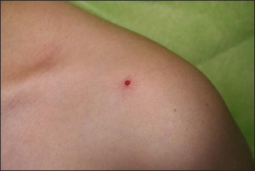
Before treatment: Spider angioma on the left upper part of the chest in a 11 year-old-boy
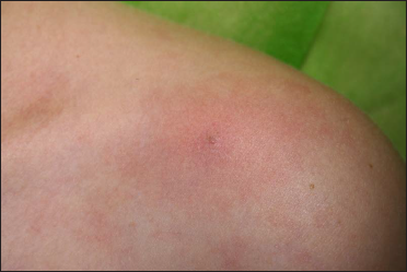
Figure 1a:
Before treatment: Spider angioma on the left upper part of the chest in a 11 year-old-boy
Immediately after copper vapour laser treatment (average power - 0.7 W, exposure time - 0.2 s)
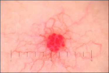
Figure 1b:
Immediately after copper vapour laser treatment (average power - 0.7 W, exposure time - 0.2 s)
Before treatment: Dermoscopy (×10) of spider angioma. The focus is symmetrical, boundaries are clear and the lobular structure is characteristic of eruptive angioma.
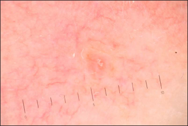
Figure 2a:
Before treatment: Dermoscopy (×10) of spider angioma. The focus is symmetrical, boundaries are clear and the lobular structure is characteristic of eruptive angioma.
Immediately after copper vapour laser treatment. Dermoscopy (×10) of spider angioma: the focus has changed and acquired a white-grey tint due to coagulated vessels and cavities.
Figure 2b:
Immediately after copper vapour laser treatment. Dermoscopy (×10) of spider angioma: the focus has changed and acquired a white-grey tint due to coagulated vessels and cavities.

Informed consent was taken after discussing the risks and benefits of laser treatment. The lesions were treated with copper vapour laser (Yakhroma–Med, Lebedev Physical Institute of Russian Academy of Sciences). The treatment was performed without anaesthesia. The laser settings were as follows: average power of 0.5–0.65 W at yellow (578 nm) wavelength, the exposure time was 0.1–0.2 s and the diameter of the light spot on the skin was 1 mm. The treatment endpoint using a multiple–stacking–pass technique was assumed when the treated area acquired a greyish tint or blanching [Figures 1b, 2b, 3b and 4b].
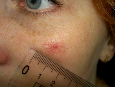
Before treatment: Spider angioma on the right cheek in a 25-year-old woman
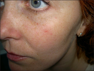
Figure 3a:
Before treatment: Spider angioma on the right cheek in a 25-year-old woman
Two weeks after copper vapour laser treatment: Spider angioma on the right cheek in a 25-year old woman
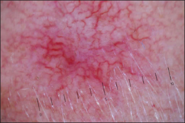
Figure 3b:
Two weeks after copper vapour laser treatment: Spider angioma on the right cheek in a 25-year old woman
Dermoscopy (×10) of spider angioma before treatment
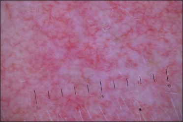
Figure 4a:
Dermoscopy (×10) of spider angioma before treatment
Dermoscopy (×10) of spider angioma after tracing the “legs” surrounding the core vessel. The focus has changed dramatically and acquired a white tint due to obliterated ectatic vessels and cavities.
Figure 4b:
Dermoscopy (×10) of spider angioma after tracing the “legs” surrounding the core vessel. The focus has changed dramatically and acquired a white tint due to obliterated ectatic vessels and cavities.

In Figure 5, we have demonstrated the spider angioma at various steps of laser treatment. Spider angioma is usually treated by tracing the “legs” surrounding the core vessel at an average power of 0.55 W; the exposure time being 0.12 s [Figure 5b]. Following, the power is increased up to 0.65 W, the exposure time increased up to 0.2 s, and the core vessel is sealed [Figure 5c]. The core vessel should be fully obliterated. Treating the radial dilated arterioles legs initially reduces the flow to the central core. The diminished blood flow to the core larger vessel contributes to complete photothermic obliteration of telangiectatic vessels.
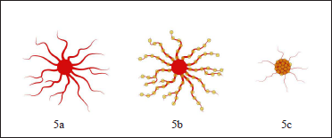
The spider angioma treatment steps: Before treatment (5a), tracing the “legs” surrounding the core vessel (5b), power increases and the core vessel is sealed (5c). Yellow spots indicate the position of the laser pulses during the treatment.
Figure 5:
The spider angioma treatment steps: Before treatment (5a), tracing the “legs” surrounding the core vessel (5b), power increases and the core vessel is sealed (5c). Yellow spots indicate the position of the laser pulses during the treatment.

The removal of spider angioma was carried out in a single session. No bleeding or erythema occurred post-treatment. After the procedure, the irradiated skin was treated with a 0.05% chlorhexidine gluconate solution and then epithelializing Bepanthen cream twice a day. Dermoscopy (×10) was used to check if there were some dilated vessels remaining after the laser irradiation. [Figures 2b and 4b].

The irradiated skin healed with crusting and exfoliation for two weeks [Figure 3b]. The treated spider angioma was evaluated 6–8 weeks after treatment. There were no recurrences for 24 months post-laser treatment. The treatment with copper vapour laser provided the complete removal of pyogenic angioma and clearance without adverse effect in any case. Copper vapour laser radiation provided complete removal of spider angioma by the appropriate vascular obliteration due to the high absorption of 578 nm radiation by oxyhaemoglobin in arterioles.

Our study demonstrates an excellent safety profile of copper vapour laser when treating spider angioma without purpura or pigmentary changes with good cosmetic results. Dermoscopy may detect subtle changes in the microvascular structure of the skin during the laser treatment that the naked eye cannot observe and thus improves the results of laser treatment to prevent relapses.

The limitation of the study is the lack of comparative assessment of the optimal copper vapour laser settings for patients with different skin phototypes.

Acknowledgement

The authors wish to thank Dr. Nataliya Melnikova, Medical Center LaNezh, Vologda, for supporting this study.

Declaration of patient consent

The authors certify that they have obtained all appropriate patient consent forms.

Financial support and sponsorship

Nil.

Conflict of interest

There are no conflicts of interest.

References

  1. , , . Spiders on the skin: Spider angioma. J Symptoms Signs. 2012;1:15.
    [Google Scholar]
  2. , , . Treatment of superficial vascular lesions with the KTP 532-nm laser: experience with 647 patients. Lasers Med Sci. 2014;29:267-71.
    [CrossRef] [PubMed] [Google Scholar]
  3. , , , . Recurrence of spider naevi following treatment with 585 nm pulsed dye laser. J Plast Reconstr Aesthet Surg. 2007;60:668-71.
    [CrossRef] [PubMed] [Google Scholar]
  4. , , , . Comparative numerical analysis and optimization of blood vessels heated using various lasers. Laser Physics. 2018;28:096003.
    [Google Scholar]
  5. , , , , . An evaluation of the copper-bromide laser for treating telangiectasia. Dermatol Surg. 1996;22:551-7.
    [CrossRef] [PubMed] [Google Scholar]
  6. , , , . Treatment of pyogenic granuloma in children with copper vapor laser radiation (578 nm) Indian J Dermatol Venereol Leprol. 2021;87:856-8.
    [CrossRef] [PubMed] [Google Scholar]
  7. , , . Numerical modeling and clinical evaluation of pulsed dye laser and copper vapor laser in skin vascular lesions treatment. J Lasers Med Sci. 2019;10:44-9.
    [CrossRef] [PubMed] [Google Scholar]
  8. , , , . Single-center experience with potassium titanyl phosphate (KTP) laser for superficial cutaneous vascular lesions in face. J Cosmet Laser Ther. 2016;18:428-31.
    [CrossRef] [PubMed] [Google Scholar]

Fulltext Views
3,497

PDF downloads
2,285
View/Download PDF
Download Citations
BibTeX
RIS
Show Sections