Translate this page into:
Triads in dermatology
Correspondence Address:
Chitra Nayak
Department of Skin & VD, OPD 14, Second floor, OPD building, Topiwala National Medical College and B.Y.L. Nair Hospital, Mumbai Central, Mumbai
India
| How to cite this article: Madke B, Nayak C. Triads in dermatology. Indian J Dermatol Venereol Leprol 2012;78:657-660 |
Most of the medical and surgical diseases/conditions/disorders are characterized by some clinical symptoms/signs/findings/observations, which form a triad and is considered pathognomonic for that condition. Signs and symptoms of various disorders are not always easy to remember. If there are three main signs and symptoms in a condition, grouping them as a triad facilitates their recall. We list here many such triads---however no explanation of the pathophysiology of these clinical signs and symptoms is undertaken as that is beyond the scope of this paper.
Adiposis dolorosa (Dercum′s disease)
This rare idiopathic condition is characterized by painful plaques, ecchymosis, and obesity, in the menopausal women. [1]
Birt-Hogg-Dube syndrome
It is an autosomal dominant condition having benign skin appendageal tumors/hamartomas that show difference toward hair follicles, i.e., (a) fibrofolliculomas (toward the mantle of the hair follicle); (b) trichodiscomas (toward the mesodermal components of the hair discs); and (c) acrochordons (skin tags). [2]
Churg-Strauss syndrome (Allergic granulomatosis)
This disease is characterized by asthma, peripheral blood eosinophilia, and necrotizing vasculitis with extravascular granulomas. [3]
Cryoglobulinemia (Meltzer′s triad)
Purpura, asthenia, and arthralgia have been together termed Meltzer′s triad seen in cryoglobulnemia. [4]
Congenital syphilis (Hutchinson′s triad)
The triad of interstitial keratitis, Hutchinson′s teeth, and eighth-nerve deafness forms "Hutchinson′s triad" of syphilitic stigmata. [5]
Dyskeratosis congenita
Reticulate hyperpigmentation of the skin, nail dystrophy, and leukokeratosis of mucous membrane. [6] [Figure - 1] and [Figure - 2]
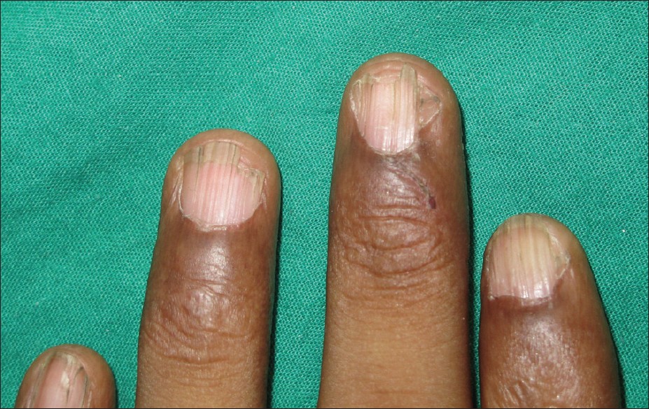 |
| Figure 1: Onychodystrophy of fingernails characterized by longitudinal ridging |
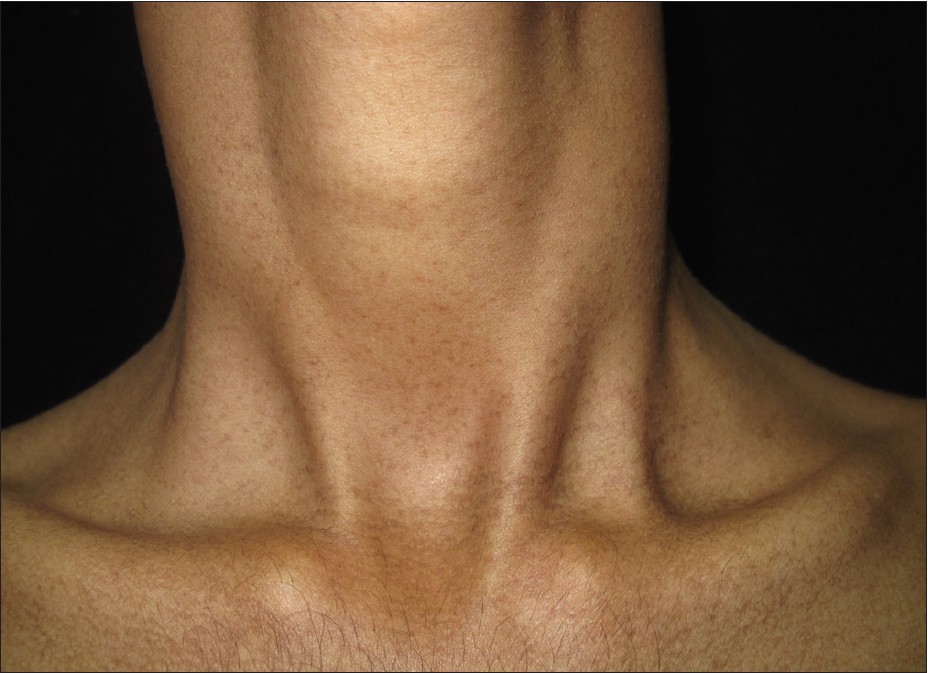 |
| Figure 2: Reticulate pattern of hyperpigmentation on anterior aspect of neck |
Follicular Occlusion Triad/Tetrad
Acne congoblata, dissecting cellulitis of scalp, pilonidal sinus, and hidradenitis suppuritiva. [7]
Gorlin Goltz syndrome (Basal cell nevus syndrome)
The autosomal dominant Patched mutation condition is characterized by (i) more than two basal cell carcinomas or one appearing in patient < 20 years old; (ii) odontogenic cyst of the jaw confirmed by histopathology; (iii) three or more palmar or plantar pits. [8]
Graham Little-Piccardi-Lassueur syndrome
Triad of cicatricial alopecia of the scalp, nonscarring alopecia of the axillae and/or groin, and keratotic follicular papules. [9]
Grave′s disease
Pretibial myxedema, thyroid acropachy, and exophthalmos-Diamond′s triad. [10]
HAIR-AN syndrome
The triad of hyperandrogenism, insulin resistance, and acanthosis nigricans in women is known as the HAIR-AN syndrome. [11]
Hermansky-Pudlak syndrome
Triad of albinism, platelets lacking dense bodies and storage of ceroid-like material in tissues. [12]
Klippel-Trenaunay syndrome
The condition is characterized by the triad of (i) capillary malformations (port-wine stain/nevus flammeus), (ii) venous malformations (abnormal varicosities, persistent embryonic veins, and (iii) disproportionate limb growth of soft tissue and/or bone [13] [Figure - 3].
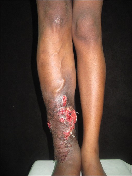 |
| Figure 3: Ateriovenous malformation of right lower limb with limb lengthening and cutaneous ulceration |
Kasabach Merritt syndrome
Triad comprising vascular tumors, thrombocytopenia, and bleeding diathesis. [14]
Livedoid vasculopathy
Triad of livedo reticularis, atrophie blanche, and very painful, small punched-out ulcers that have a very poor tendency for healing [15] [Figure - 4].
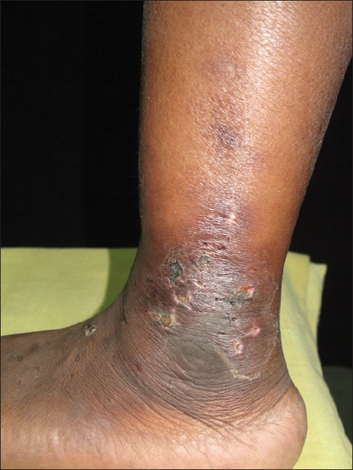 |
| Figure 4: Multiple punched ulcers on lateral malleolar area with porcelain white scars |
McCune Albright syndrome
The condition is characterized by triad of poly/monostotic fibrous dysplasia, café-au-lait macules (CALMs), and hyperfunctioning endocrinopathies including precocious puberty, hyperthyroidism, and hypercortisolism, hypersomatotropism, and hypophosphatemic rickets. [16]
Melkersson-Rosenthal syndrome
Triad of recurrent labial and/or recurrent facial edema, relapsing facial paralysis, and fissured tongue [17] [Figure - 5].
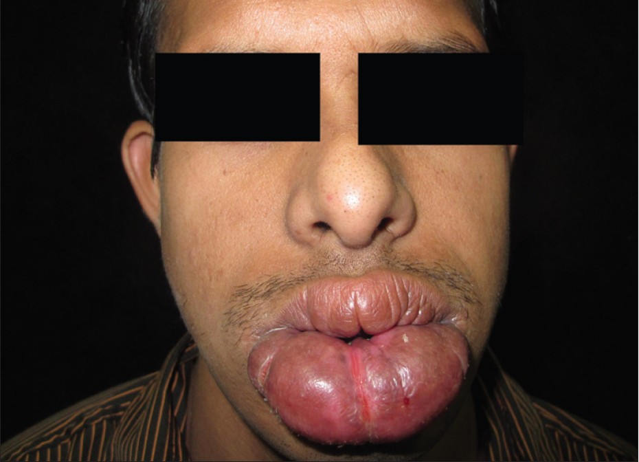 |
| Figure 5: Gross diffuse swelling of lower lip with fissuring at places |
Naxos syndrome
This autosomal condition is due to desmoplakin mutation known for its triad of woolly hair, striate palmar-plantar keratoderma, and arrythmogenic right ventricular cardiomyopathy. [18]
Netherton-Comel syndrome
Netherton Comel syndrome has two triads. This autosomal recessive ichthyosiform disorder is typified by triad of generalized infantile erythroderma, diarrhea, and failure to thrive from the perspective of a general physician. Dermatologic examination reveals a classical clinical triad of ichthyosis linearis circumflexa, trichorrhexis invaginata (bamboo hair), and atopic diathesis. [19]
Pellagra
This niacin deficiency disorder is characterized by clinical triad of 3 Ds i.e., dermatitis, diarrhea, and dementia and rarely by the fourth ′D′-- death. [20]
Primary systemic amyloidosis
Triad of carpal tunnel syndrome, macroglossia, and mucocutaneous skin lesions. [21]
Ramsay--Hunt syndrome (Herpes zoster oticus)
As the name suggests VZV infection of the facial nerve with ipsilateral facial palsy along with ear pain and vesicles on external ear or tympanic membrane constitutes the triad of Ramsay--Hunt syndrome. [22]
Reiter′s syndrome
The triad of urethritis, arthritis, and conjunctivitis forms the triad of reactive arthritis or Reiter′s syndrome. [23]
Sèzary syndrome
Sèzary syndrome is characterized by triad of (i) erythroderma, (ii) peripheral lymphadenopathy, and (iii) atypical mononuclear cells (Sézary cells) comprising 5% or more of peripheral blood lymphocytes on a buffy coat smear, or more than 20% of total lymphocyte count or a total Sézary count of more than 1000/mm 3 . [24]
Sclero-Atrophic syndrome of Huriez
Autosomal dominant congenital syndrome comprises the triad of diffuse sclero-atrophy of the dorsal aspect of the hands; palmoplantar keratoderma and hypoplastic nail changes. The characteristic electron microscopic picture shows the absence of epidermal Langerhans cells contributing to increased incidence of malignancy. [25]
Tuberous sclerosis: Vogt′s Triad (epiloia)
This is described for a neurocutaneous disorder, tuberous sclerosis characterized by diagnostic clinical triad of epilepsy, low intelligence, and facial angiofibromas (adenoma sebaceum). [26]
Wegener′s granulomatosis
It is classically described as a triad consisting of systemic small vessel vasculitis, necrotizing granulomatous inflammation of both the upper and lower respiratory tracts, and glomerulonephritis. [27]
Wiskott-Aldrich syndrome
Triad of early onset thrombocytopenia, recurrent infection, and eczema characterizes this X-linked recessive condition. [28]
Yellow nail syndrome
Yellow nail syndrome is the triad of yellow nails, primary lymphedema, and pleural effusion. [29]
The above list of triads may increase in future as many more are to be added to it. For now, students of dermatology will find this list helpful especially while revising for their examinations.
| 1. |
Chopra A, Walia P, Chopra D, Jassal JS. Adiposis dolorosa. Indian J Dermatol Venereol Leprol 2000;66:101-2.
[Google Scholar]
|
| 2. |
Collins GL, Somach S, Morgan MB. Histomorphologic and immunophenotypic analysis of fibrofolliculomas and trichodiscomas in Birt-Hogg-Dube syndrome and sporadic disease. J Cutan Pathol 2002;29:529-33.
[Google Scholar]
|
| 3. |
Choi JH, Ahn IS, Lee HB, Park CW, Lee CH, Ahn HK. A case of churg-strauss syndrome. Ann Dermatol 2009;21:213-6.
[Google Scholar]
|
| 4. |
Motyckova G, Murali M. Laboratory testing for cryoglobulins. Am J Hematol 2011;86:500-2.
[Google Scholar]
|
| 5. |
Singhal P, Patel P, Marfatia YS. A case of congenital syphilis with Hutchinson's triad. Indian J Sex Transm Dis 2011;32:34-6.
[Google Scholar]
|
| 6. |
Gupta V, Kumar A. Dyskeratosis congenital. Adv Exp Med Biol 2010;685:215-9.
[Google Scholar]
|
| 7. |
Scheinfeld NS. A case of dissecting cellulitis and a review of the literature. Dermatol Online J 2003;9:8.
[Google Scholar]
|
| 8. |
Sirous M, Tayari N. A case report of Gorlin-Goltz syndrome as a rare hereditary disorder. J Res Med Sci 2011;16:836-40.
[Google Scholar]
|
| 9. |
Vashi N, Newlove T, Chu J, Patel R, Stein J. Graham Little-Piccardi-Lassueur syndrome. Dermatol Online J 2011;17:30-1.
[Google Scholar]
|
| 10. |
Kashkouli MB, Kaghazkanani R, Heidari I, Ketabi N, Jam S, Azarnia S, et al. Bilateral versus unilateral thyroid eye disease. Indian J Ophthalmol 2011;59:363-6.
[Google Scholar]
|
| 11. |
Elmer KB, George RM. HAIR-AN syndrome: A multisystem challenge. Am Fam Physician 2001;63:2385-90.
[Google Scholar]
|
| 12. |
Sandrock K, Zieger B. Current strategies in diagnosis of inherited storage pool defects. Transfus Med Hemother 2010;37:248-58.
[Google Scholar]
|
| 13. |
Zea MI, Hanif M, Habib M, Ansari A. Klippel-Trenaunay Syndrome: A case report with brief review of literature. J Dermatol Case Rep 2009;3:56-9.
[Google Scholar]
|
| 14. |
Madke B, Doshi B, Pande S, Khopkar U. Phenomena in dermatology. Indian J Dermatol Venereol Leprol 2011;77:264- 75.
[Google Scholar]
|
| 15. |
Tabata N, Oonami K, Ishibashi M, Yamazaki M. Livedo vasculopathy associated with IgM anti-phosphatidylserine-prothrombin complex antibody. Acta Derm Venereol 2010;90:313-4.
[Google Scholar]
|
| 16. |
Saggini A, Brandi ML. Skin lesions in hereditary endocrine tumor syndromes. Endocr Pract 2011;17(Suppl 3):47-57.
[Google Scholar]
|
| 17. |
van der Waal RI, Schulten EA, van der Meij EH, van de Scheur MR, Starink TM, van der Waal I. Cheilitis granulomatosa: Overview of 13 patients with long-term follow-up: Results of management. Int J Dermatol 2002;41:225-9.
[Google Scholar]
|
| 18. |
Meera G, Prabhavathy D, Jayakumar S, Tharini G. Naxos disease in two siblings. Int J Trichology 2010;2:53-5.
[Google Scholar]
|
| 19. |
Rakowska A, Kowalska-Oledzka E, Slowinska M, Rosinska D, Rudnicka L. Hair shaft videodermoscopy in Netherton syndrome. Pediatr Dermatol 2009;26:320-2.
[Google Scholar]
|
| 20. |
Kaimal S, Thappa DM. Diet in dermatology: Revisited. Indian J Dermatol Venereol Leprol 2010;76:103-15.
[Google Scholar]
|
| 21. |
Saoji V, Chaudhari S, Gohokar D. Primary systemic amyloidosis: Three different presentations. Indian J Dermatol Venereol Leprol 2009;75:394-7.
[Google Scholar]
|
| 22. |
Sobn AJ, Tranmer PA. Ramsay Hunt syndrome in a patient with human immunodeficiency virus infection. J Am Board Fam Pract 2001;14:392-4.
[Google Scholar]
|
| 23. |
Chun P, Kim YJ, Han YM, Kim YM. A case of reactive arthritis after Salmonella enteritis in a 12-year-old boy. Korean J Pediatr 2011;54:313-5.
[Google Scholar]
|
| 24. |
Möbs M, Knott M, Fritzen B, Pullmann S, Sterry W, Assaf C. Diagnostic tools in Sezary syndrome. G Ital Dermatol Venereol 2010;145:385-91.
[Google Scholar]
|
| 25. |
Downs AM, Kennedy CT. Scleroatrophic syndrome of Huriez in an infant. Pediatr Dermatol 1998;15:207-9.
[Google Scholar]
|
| 26. |
Purkait R, Samanta T, Thakur S, Dhar S. Neurocutaneous syndrome: A prospective study. Indian J Dermatol 2011;56:375- 9.
[Google Scholar]
|
| 27. |
Mahajan V, Whig J, Kashyap A, Gupta S. Diffuse alveolar hemorrhage in Wegener's granulomatosis. Lung India 2011;28:52-5.
[Google Scholar]
|
| 28. |
Syrigos KN, Makrilia N, Neidhart J, Moutsos M, Tsimpoukis S, Kiagia M, et al. Prolonged survival after splenectomy in Wiskott-Aldrich syndrome: A case report. Ital J Pediatr 2011;37:42.
[Google Scholar]
|
| 29. |
Avitan-Hersh E, Berger G, Bergman R. Yellow nail syndrome. Isr Med Assoc J 2011;13:577-8.
[Google Scholar]
|
Fulltext Views
8,862
PDF downloads
5,999


