Generic selectors
Exact matches only
Search in title
Search in content
Post Type Selectors
Search in posts
Search in pages
Filter by Categories
15th National Conference of the IAOMFP, Chennai, 2006
Abstract
Abstracts from current literature
Acne in India: Guidelines for management - IAA Consensus Document
Addendum
Announcement
Art & Psychiatry
Article
Articles
Association Activities
Association Notes
Award Article
Book Review
Brief Report
Case Analysis
Case Letter
Case Letters
Case Notes
Case Report
Case Reports
Clinical and Laboratory Investigations
Clinical Article
Clinical Studies
Clinical Study
Commentary
Conference Oration
Conference Summary
Continuing Medical Education
Correspondence
Corrigendum
Cosmetic Dermatology
Cosmetology
Current Best Evidence
Current Issue
Current View
Derma Quest
Dermato Surgery
Dermatopathology
Dermatosurgery Specials
Dispensing Pearl
Do you know?
Drug Dialogues
e-IJDVL
Editor Speaks
Editorial
Editorial Remarks
Editorial Report
Editorial Report - 2007
Editorial report for 2004-2005
Errata
Erratum
Focus
Fourth All India Conference Programme
From Our Book Shelf
From the Desk of Chief Editor
General
Get Set for Net
Get set for the net
Guest Article
Guest Editorial
History
How I Manage?
IADVL Announcement
IADVL Announcements
IJDVL Awards
IJDVL AWARDS 2015
IJDVL Awards 2018
IJDVL Awards 2019
IJDVL Awards 2020
IJDVL International Awards 2018
Images in Clinical Practice
Images in Dermatology
In Memorium
Inaugural Address
Index
Knowledge From World Contemporaries
Leprosy Section
Letter in Response to Previous Publication
Letter to Editor
Letter to the Editor
Letter to the Editor - Case Letter
Letter to the Editor - Letter in Response to Published Article
LETTER TO THE EDITOR - LETTERS IN RESPONSE TO PUBLISHED ARTICLES
Letter to the Editor - Observation Letter
Letter to the Editor - Study Letter
Letter to the Editor - Therapy Letter
Letter to the Editor: Articles in Response to Previously Published Articles
Letters in Response to Previous Publication
Letters to the Editor
Letters to the Editor - Letter in Response to Previously Published Articles
Letters to the Editor: Case Letters
Letters to the Editor: Letters in Response to Previously Published Articles
Media and news
Medicolegal Window
Messages
Miscellaneous Letter
Musings
Net Case
Net case report
Net Image
Net Images
Net Letter
Net Quiz
Net Study
New Preparations
News
News & Views
Obituary
Observation Letter
Observation Letters
Oration
Original Article
ORIGINAL CONTRIBUTION
Original Contributions
Pattern of Skin Diseases
Pearls
Pediatric Dermatology
Pediatric Rounds
Perspective
Presedential Address
Presidential Address
Presidents Remarks
Quiz
Recommendations
Regret
Report
Report of chief editor
Report of Hon : Treasurer IADVL
Report of Hon. General Secretary IADVL
Research Methdology
Research Methodology
Resident page
Resident's Page
Resident’s Page
Residents' Corner
Residents' Corner
Residents' Page
Retraction
Review
Review Article
Review Articles
Reviewers 2022
Revision Corner
Self Assessment Programme
SEMINAR
Seminar: Chronic Arsenicosis in India
Seminar: HIV Infection
Short Communication
Short Communications
Short Report
Snippets
Special Article
Specialty Interface
Studies
Study Letter
Study Letters
Supplement-Photoprotection
Supplement-Psoriasis
Symposium - Contact Dermatitis
Symposium - Lasers
Symposium - Pediatric Dermatoses
Symposium - Psoriasis
Symposium - Vesicobullous Disorders
SYMPOSIUM - VITILIGO
Symposium Aesthetic Surgery
Symposium Dermatopathology
Symposium-Hair Disorders
Symposium-Nails Part I
Symposium-Nails-Part II
Systematic Review and Meta-Analysis
Systematic Reviews and Meta-analyses
Systematic Reviews and Meta-analysis
Tables
Technology
Therapeutic Guideline-IADVL
Therapeutic Guidelines
Therapeutic Guidelines - IADVL
Therapeutics
Therapy
Therapy Letter
Therapy Letters
View Point
Viewpoint
What’s new in Dermatology
View/Download PDF

Translate this page into:

Case Letter
88 (
4
); 536-540
doi:
10.25259/IJDVL_7_2020
pmid:
35593282

Unilateral areolar leiomyoma with sebaceous hyperplasia

Department of Dermatology, University of Tsukuba, Tsukuba, Ibaraki, Japan
1-1-1 Tennodai, Tsukuba, Japan

Corresponding author: Dr. Yoshiyuki Nakamura, Department of Dermatology, University of Tsukuba, Tsukuba, Ibaraki, Japan, ynakamura-tuk@umin.ac.jp

Licence
This is an open-access article distributed under the terms of the Creative Commons Attribution-Non Commercial-Share Alike 4.0 License, which allows others to remix, transform, and build upon the work non-commercially, as long as the author is credited and the new creations are licensed under the identical terms.

How to cite this article: Oya K, Nakamura Y, Fujisawa Y. Unilateral areolar leiomyoma with sebaceous hyperplasia. Indian J Dermatol Venereol Leprol 2022;88:536-40.

Sir,

Leiomyoma frequently develops in the uterus, but its occurrence in the areola is rare. Similarly, sebaceous hyperplasia, usually located on the nose and forehead, also rarely occurs in the areola. However, cases where both leiomyoma and sebaceous hyperplasia developed in the same areolar lesion have not been reported. Here, we report such a case in the unilateral areola.

A 41-year-old man noticed a nodule on his left areola one year before his initial visit to our hospital. On physical examination, a 1-cm, yellowish nodule was found on the upper part of the left areola [Figures 1a and b]. Polarized light dermatoscopy (Heine Delta 20 plus; Heine Optotechnik, Herrsching, Germany) revealed white reticular lines with yellowish globular structures [Figure 1c] and a biopsy was performed. Histopathology of the lesion showed numerous, mature sebaceous glands whose ducts directly led to the epidermis [Figures 1d and e]. In addition, moderate epidermal hyperplasia with elongated rete ridges was also observed [Figures 1d and e] and, in the dermis, tumour nests composed of α-smooth muscle actin-positive spindle-shaped cells with intermingled collagen were seen [Figures 1d, f and Figure 2a]. These results led to a diagnosis of areolar leiomyoma with sebaceous hyperplasia. Additional immunohistochemical studies revealed that the leiomyoma was positive for estrogen and progesterone receptors but the proliferated sebocytes were negative [Figures 2b and c]. In contrast, the sebocytes were positive for androgen receptors but the leiomyoma was negative [Figure 2d]. Epidermal growth factor, insulin-like growth factor-1 and fibroblast growth factor-2 were highly expressed in the leiomyoma [Figures 3a-c and 4a-d]. Epidermal growth factor receptor, insulin-like growth factor-1 receptor β and fibroblast growth factor receptor 2, a receptor for fibroblast growth factor-2, were expressed in the leiomyoma, the sebocytes and the adjacent keratinocytes [Figures 3d-i and 4e-h]. In addition, phosphorylated extracellular signal-regulated kinase 1/2 and phosphorylated Akt were positive in the leiomyoma, the sebocytes and the adjacent keratinocytes [Figures 3j-m and 4a, e, i and j]. Although the patient refused resection of the whole lesion, it has remained stable for more than one year.

Smooth muscle hamartoma should be considered for the differential diagnosis of dermal lesions such as the lesion of the present case. This disease generally presents as variably sized papules, patches, and plaques. Histologically, this disease comprises variably oriented, discrete, and well-defined bundles of smooth muscle cells. These clinicopathologic findings differ from those of the present case, which presented clinically as a nodule and with histologic findings of smooth muscle cell proliferation with intermingled collagen, findings consistent with leiomyoma. Cutaneous leiomyomas are categorized into pilar, angio- and genital leiomyomas and areolar leiomyoma is a rare variant of genital leiomyoma. They may show overlying epidermal hyperplasia as frequently seen in pilar leiomyoma although the mechanism of this remains elusive.

Some growth factors such as epidermal growth factor, insulin-like growth factor and fibroblast growth factor may play crucial roles for sebocyte proliferation.1 In addition, some dermal tumours, such as dermatofibroma, are thought to induce epithelial hyperplasia through growth factor secretion.2 Uterine leiomyoma has also been reported to produce some growth factors, which may promote the growth of the tumour cells in an autocrine manner.3 Moreover, endometrial hyperplasia adjacent to uterine leiomyoma is often seen.4 This may be due to the hyperestrogenic status. However, given that growth factors play important roles in endometrial proliferation, growth factors produced from uterine leiomyoma might also contribute to endometrial hyperplasia via paracrine action.5 In the present case, tumour cells of the leiomyoma showed high expression levels of epidermal growth factor, insulin-like growth factor-1 and fibroblast growth factor-2. These receptors were also expressed in the leiomyoma. In addition, phosphorylated extracellular signal-regulated kinase 1/2 and phosphorylated Akt, representative markers of the activated downstream signalling pathways of the growth factor receptors, were positive in the leiomyoma, suggesting that growth factors from the leiomyoma might have promoted the growth of the leiomyoma itself in an autocrine manner identical to uterine leiomyoma. Moreover, in the present case, the sebocytes and the adjacent keratinocytes expressed epidermal growth factor receptor, insulin-like growth factor-1 receptor β and fibroblast growth factor receptor 2 while also being positive for phosphorylated extracellular signal-regulated kinase 1/2 and phosphorylated Akt, suggesting that the sebaceous hyperplasia with moderate acanthosis might have developed due to the growth factors produced by the underlying leiomyoma.

In conclusion, we describe a case where both leiomyoma and sebaceous hyperplasia were observed in the unilateral areola. Although the exact pathogenesis of the rare association in the present case remains unknown, we speculate that the leiomyoma-produced growth factors including epidermal growth factor, insulin-like growth factor-1 and fibroblast growth factor-2 may have been involved in the growth of the leiomyoma itself in an autocrine manner and of the overlying sebocytes and keratinocytes by paracrine action.

A 1 cm nodule (black arrow) was observed on the left areola
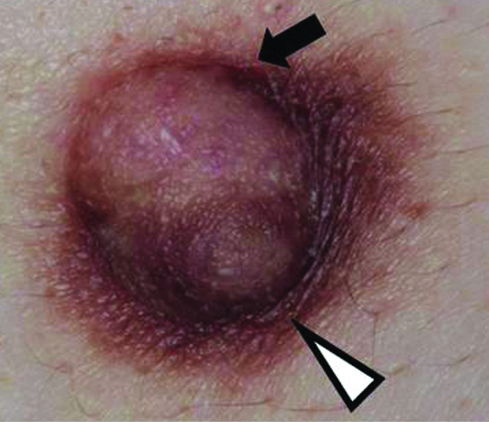
Figure 1a:
A 1 cm nodule (black arrow) was observed on the left areola
The white arrowhead and black arrow indicate the left nipple and nodule, respectively
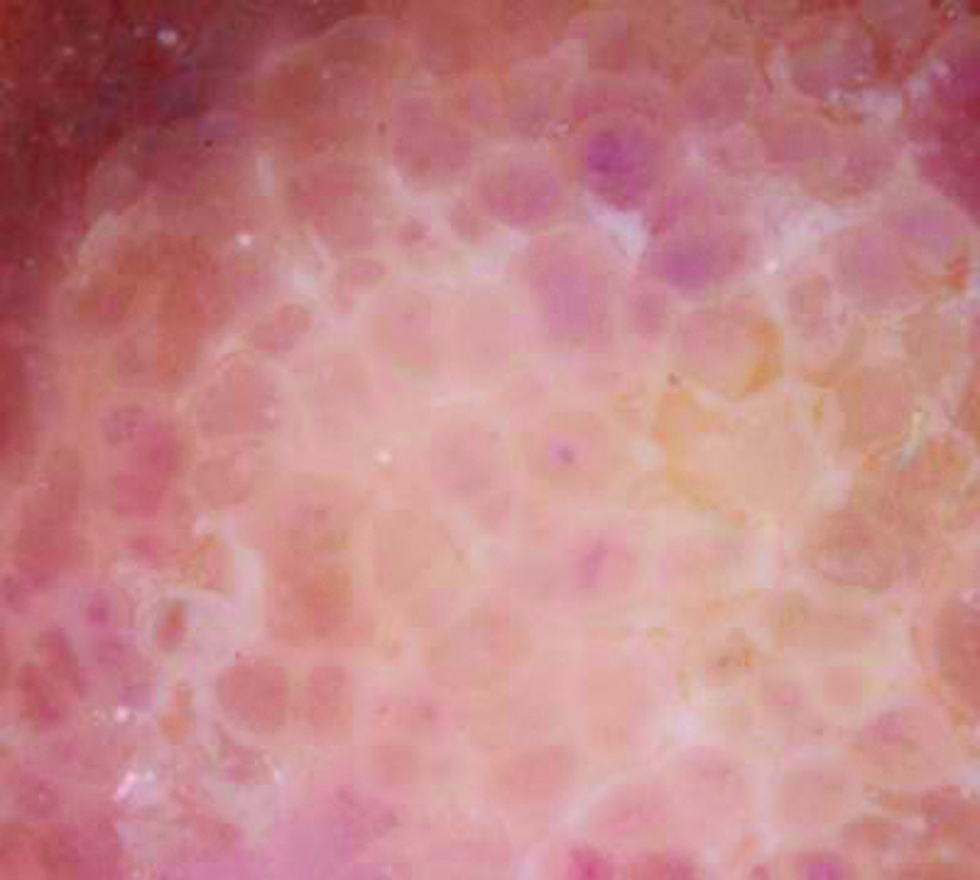
Figure 1b:
The white arrowhead and black arrow indicate the left nipple and nodule, respectively
White reticular lines with yellowish globules (polarized, ×10)
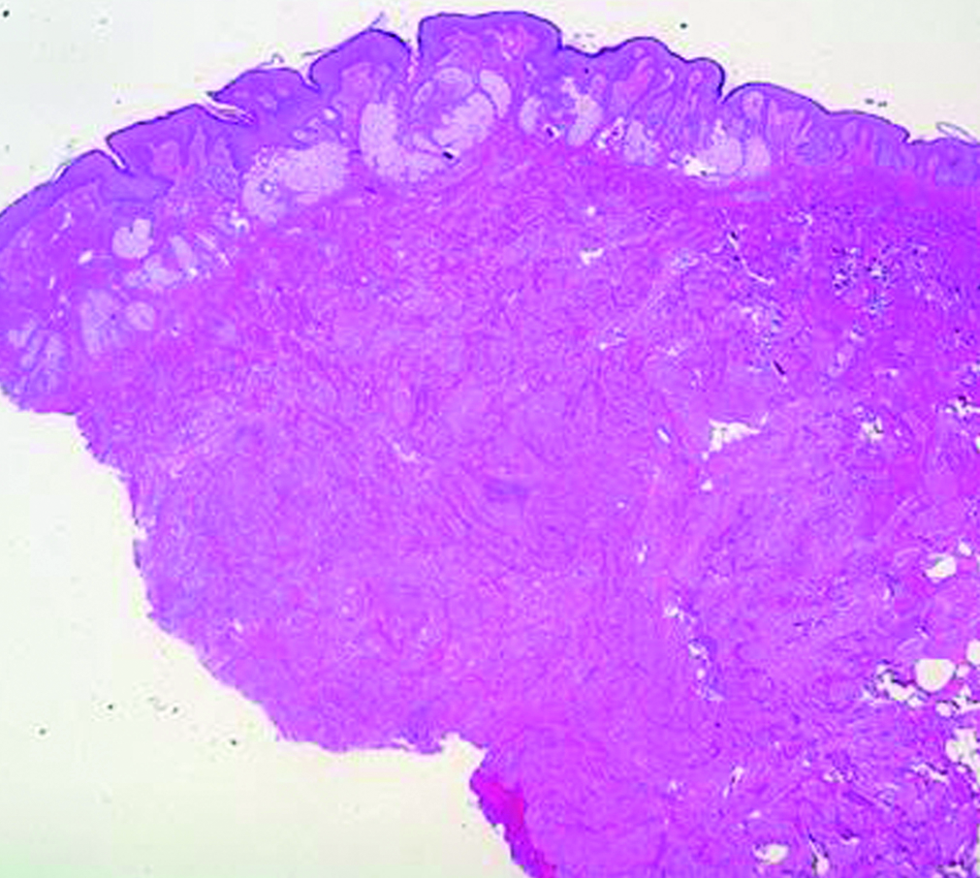
Figure 1c:
White reticular lines with yellowish globules (polarized, ×10)
Large numbers of mature sebaceous glands and dermal tumour nests visible in the dermis. (Hematoxylin and eosin, ×20)
Figure 1d:
Large numbers of mature sebaceous glands and dermal tumour nests visible in the dermis. (Hematoxylin and eosin, ×20)
The proliferated sebaceous glands leading directly to the epidermis. Moderate acanthosis with elongated rete ridges are also visible (Haematoxylin and eosin, ×100)
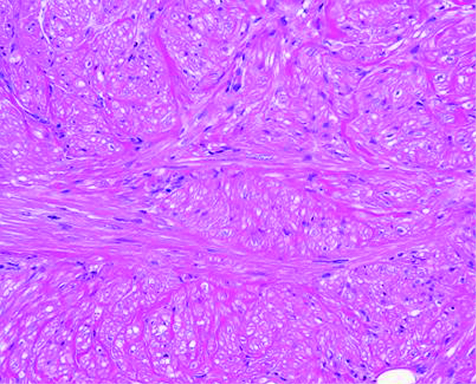
Figure 1e:
The proliferated sebaceous glands leading directly to the epidermis. Moderate acanthosis with elongated rete ridges are also visible (Haematoxylin and eosin, ×100)
The dermal tumour cells with a spindle-shaped morphology (Haematoxylin and eosin, ×200)
Figure 1f:
The dermal tumour cells with a spindle-shaped morphology (Haematoxylin and eosin, ×200)
The dermal tumour cells showing positive staining for (a) α-smooth muscle actin (×200), (b) estrogen receptors (×400) and (c) progesterone receptors (×400); (d) Proliferated sebocytes testing positive for androgen receptors (×200)
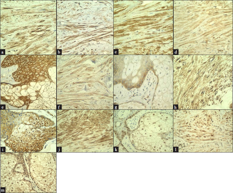
Figure 2:
The dermal tumour cells showing positive staining for (a) α-smooth muscle actin (×200), (b) estrogen receptors (×400) and (c) progesterone receptors (×400); (d) Proliferated sebocytes testing positive for androgen receptors (×200)
High expression in the leiomyoma displayed by (a) epidermal growth factor (×400), (b) insulin-like growth factor-1 (×400) and (c) fibroblast growth factor-2 (×400); (d) Epidermal growth factor receptor showing expression in the leiomyoma (×400), (e) Epidermal growth factor receptor showing expression in the sebocytes with the adjacent keratinocytes (×400), (f) Insulin-like growth factor-1 receptor β showing expression in the leiomyoma (×400), (g) Insulin-like growth factor-1 receptor β showing expression in the sebocytes with the adjacent keratinocytes (×400), (h) Fibroblast growth factor receptor 2 showing expression in the leiomyoma (×400), (i) Fibroblast growth factor receptor 2 showing expression in the sebocytes with the adjacent keratinocytes (×400), (j) Positive staining of phosphorylated extracellular signal-regulated kinase 1/2 in the leiomyoma (×400), (k) Positive staining of phosphorylated extracellular signal-regulated kinase 1/2 in the sebocytes with adjacent keratinocytes (×400), (l) Positive staining of phosphorylated Akt in the leiomyoma (×400), (m) Positive staining of phosphorylated Akt in the sebocytes with adjacent keratinocytes (×400).
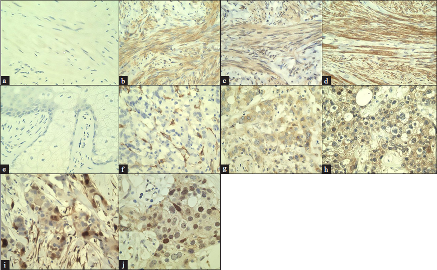
Figure 3:
High expression in the leiomyoma displayed by (a) epidermal growth factor (×400), (b) insulin-like growth factor-1 (×400) and (c) fibroblast growth factor-2 (×400); (d) Epidermal growth factor receptor showing expression in the leiomyoma (×400), (e) Epidermal growth factor receptor showing expression in the sebocytes with the adjacent keratinocytes (×400), (f) Insulin-like growth factor-1 receptor β showing expression in the leiomyoma (×400), (g) Insulin-like growth factor-1 receptor β showing expression in the sebocytes with the adjacent keratinocytes (×400), (h) Fibroblast growth factor receptor 2 showing expression in the leiomyoma (×400), (i) Fibroblast growth factor receptor 2 showing expression in the sebocytes with the adjacent keratinocytes (×400), (j) Positive staining of phosphorylated extracellular signal-regulated kinase 1/2 in the leiomyoma (×400), (k) Positive staining of phosphorylated extracellular signal-regulated kinase 1/2 in the sebocytes with adjacent keratinocytes (×400), (l) Positive staining of phosphorylated Akt in the leiomyoma (×400), (m) Positive staining of phosphorylated Akt in the sebocytes with adjacent keratinocytes (×400).
(a) Negative control staining in the leiomyoma using rabbit IgG isotype control antibody (×400); Positive control staining using a uterine leiomyoma sample for: (b) epidermal growth factor (×400), (c) insulin-like growth factor-1 (×400), (d) fibroblast growth factor-2 (×400), (e) Negative control staining in the sebocytes with adjacent keratinocytes using rabbit IgG isotype control antibody (×400); Positive control staining using a breast cancer sample for: (f) epidermal growth factor receptor (×400), (g) insulin-like growth factor-1 receptor β (×400), (h) fibroblast growth factor receptor 2 (×400), (i) phosphorylated extracellular signal-regulated kinase 1/2 (×400) and (j) phosphorylated Akt (×400)
Figure 4:
(a) Negative control staining in the leiomyoma using rabbit IgG isotype control antibody (×400); Positive control staining using a uterine leiomyoma sample for: (b) epidermal growth factor (×400), (c) insulin-like growth factor-1 (×400), (d) fibroblast growth factor-2 (×400), (e) Negative control staining in the sebocytes with adjacent keratinocytes using rabbit IgG isotype control antibody (×400); Positive control staining using a breast cancer sample for: (f) epidermal growth factor receptor (×400), (g) insulin-like growth factor-1 receptor β (×400), (h) fibroblast growth factor receptor 2 (×400), (i) phosphorylated extracellular signal-regulated kinase 1/2 (×400) and (j) phosphorylated Akt (×400)

Declaration of patient consent

Patient’s consent is not required as the patient’s identity is not disclosed or compromised.

Financial support and sponsorship

Nil.

Conflicts of interest

There are no conflicts of interest.

References

  1. , , , , , . Homeostasis of the sebaceous gland and mechanisms of acne pathogenesis. Br J Dermatol. 2019;181:677-90.
    [CrossRef] [PubMed] [Google Scholar]
  2. , , . Epithelial induction in dermatofibroma: A role for the epidermal growth factor (EGF) receptor. Am J Dermatopathol. 1997;19:35-40.
    [CrossRef] [PubMed] [Google Scholar]
  3. , . Interactions of cytokines, growth factors, and the extracellular matrix in the cellular biology of uterine leiomyomata. Fertil Steril. 2002;78:1-12.
    [CrossRef] [PubMed] [Google Scholar]
  4. , , , . Uterine Leiomyomas: An ENIGMA. J Midlife Health. 2016;7:22-7.
    [CrossRef] [PubMed] [Google Scholar]
  5. , , , , . Developmental reprogramming of IGF signaling and susceptibility to endometrial hyperplasia in the rat. Lab Invest. 2008;88:615-26.
    [CrossRef] [PubMed] [Google Scholar]

Fulltext Views
5,981

PDF downloads
2,553
View/Download PDF
Download Citations
BibTeX
RIS
Show Sections