Generic selectors
Exact matches only
Search in title
Search in content
Post Type Selectors
Search in posts
Search in pages
Filter by Categories
15th National Conference of the IAOMFP, Chennai, 2006
Abstract
Abstracts from current literature
Acne in India: Guidelines for management - IAA Consensus Document
Addendum
Announcement
Art & Psychiatry
Article
Articles
Association Activities
Association Notes
Award Article
Book Review
Brief Report
Case Analysis
Case Letter
Case Letters
Case Notes
Case Report
Case Reports
Clinical and Laboratory Investigations
Clinical Article
Clinical Studies
Clinical Study
Commentary
Conference Oration
Conference Summary
Continuing Medical Education
Correspondence
Corrigendum
Cosmetic Dermatology
Cosmetology
Current Best Evidence
Current Issue
Current View
Derma Quest
Dermato Surgery
Dermatopathology
Dermatosurgery Specials
Dispensing Pearl
Do you know?
Drug Dialogues
e-IJDVL
Editor Speaks
Editorial
Editorial Remarks
Editorial Report
Editorial Report - 2007
Editorial report for 2004-2005
Errata
Erratum
Focus
Fourth All India Conference Programme
From Our Book Shelf
From the Desk of Chief Editor
General
Get Set for Net
Get set for the net
Guest Article
Guest Editorial
History
How I Manage?
IADVL Announcement
IADVL Announcements
IJDVL Awards
IJDVL AWARDS 2015
IJDVL Awards 2018
IJDVL Awards 2019
IJDVL Awards 2020
IJDVL International Awards 2018
Images in Clinical Practice
In Memorium
Inaugural Address
Index
Knowledge From World Contemporaries
Leprosy Section
Letter in Response to Previous Publication
Letter to Editor
Letter to the Editor
Letter to the Editor - Case Letter
Letter to the Editor - Letter in Response to Published Article
LETTER TO THE EDITOR - LETTERS IN RESPONSE TO PUBLISHED ARTICLES
Letter to the Editor - Observation Letter
Letter to the Editor - Study Letter
Letter to the Editor - Therapy Letter
Letter to the Editor: Articles in Response to Previously Published Articles
Letters in Response to Previous Publication
Letters to the Editor
Letters to the Editor - Letter in Response to Previously Published Articles
Letters to the Editor: Case Letters
Letters to the Editor: Letters in Response to Previously Published Articles
Medicolegal Window
Messages
Miscellaneous Letter
Musings
Net Case
Net case report
Net Image
Net Images
Net Letter
Net Quiz
Net Study
New Preparations
News
News & Views
Obituary
Observation Letter
Observation Letters
Oration
Original Article
ORIGINAL CONTRIBUTION
Original Contributions
Pattern of Skin Diseases
Pearls
Pediatric Dermatology
Pediatric Rounds
Perspective
Presedential Address
Presidential Address
Presidents Remarks
Quiz
Recommendations
Regret
Report
Report of chief editor
Report of Hon : Treasurer IADVL
Report of Hon. General Secretary IADVL
Research Methdology
Research Methodology
Resident page
Resident's Page
Resident’s Page
Residents' Corner
Residents' Corner
Residents' Page
Retraction
Review
Review Article
Review Articles
Reviewers 2022
Revision Corner
Self Assessment Programme
SEMINAR
Seminar: Chronic Arsenicosis in India
Seminar: HIV Infection
Short Communication
Short Communications
Short Report
Snippets
Special Article
Specialty Interface
Studies
Study Letter
Study Letters
Supplement-Photoprotection
Supplement-Psoriasis
Symposium - Contact Dermatitis
Symposium - Lasers
Symposium - Pediatric Dermatoses
Symposium - Psoriasis
Symposium - Vesicobullous Disorders
SYMPOSIUM - VITILIGO
Symposium Aesthetic Surgery
Symposium Dermatopathology
Symposium-Hair Disorders
Symposium-Nails Part I
Symposium-Nails-Part II
Systematic Review and Meta-Analysis
Systematic Reviews and Meta-analyses
Systematic Reviews and Meta-analysis
Tables
Technology
Therapeutic Guideline-IADVL
Therapeutic Guidelines
Therapeutic Guidelines - IADVL
Therapeutics
Therapy
Therapy Letter
Therapy Letters
View Point
Viewpoint
What’s new in Dermatology
View/Download PDF

Translate this page into:

Net Quiz
89 (
6
); 928-928
doi:
10.25259/IJDVL_1142_2022
pmid:
37317719

Unilateral erythematous nodule over eyelid

Department of Dermatology, Seth Jagannath Pahadiya Medical College, Bharatpur, Rajasthan, India
Department of Dermatology, AIIMS Jodhpur, Rajasthan, India
Department of Pathology and Lab Medicine, AIIMS Jodhpur, Rajasthan, India
Corresponding author: Dr Anupama Bains, Department of Dermatology, AIIMS Jodhpur, Rajasthan, India. whiteangel2387@gmail.com
Licence
This is an open-access article distributed under the terms of the Creative Commons Attribution-Non Commercial-Share Alike 4.0 License, which allows others to remix, transform, and build upon the work non-commercially, as long as the author is credited and the new creations are licensed under the identical terms.

How to cite this article: Ansari F, Agrawal A, Bains A, Elhence P. Unilateral erythematous nodule over eyelid. Indian J Dermatol Venereol Leprol 2023;89:928

A 60-year-old woman from West Rajasthan presented with the complaint of an insidious onset of gradually progressive reddish swelling on the left upper eyelid for 1.5 months, which was associated with heaviness and mild pain. She denied history of insect bite, ocular symptoms, fever or any other systemic complaints. On examination, a single, well-defined, soft, non-tender, erythematous nodulo-plaque of 2 × 2 cm size was present on the left upper eyelid with overlying whitish scales and no discharge [Figure 1]. Rest of the cutaneous and systemic examination was normal. Ophthalmological evaluation was unremarkable. Ultrasonography showed a hypoechoic lesion in the subcutaneous plane. Slit skin smear for Mycobacterium leprae was negative. Punch biopsy revealed mild focal spongiosis and follicular plugging in epidermis. Grenz zone was noted. Entire dermis and subcutis showed diffuse infiltrate of epithelioid histiocytes admixed with plasma cells and lymphocytes. Histiocytes were studded with small intra-cytoplasmic, basophilic inclusion bodies which were highlighted by Giemsa stain [Figures 2a and 2b].

Figure 1
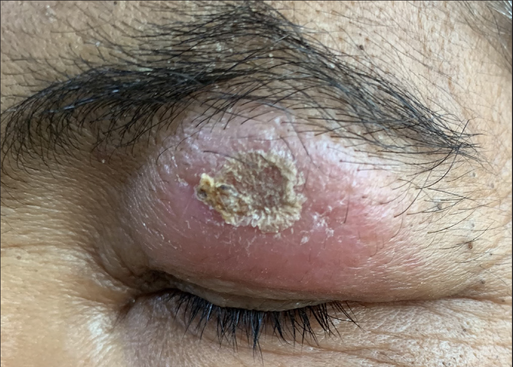
Solitary mildly erythematous scaly nodulo-plaque on the left upper eyelid.
Figure 2a
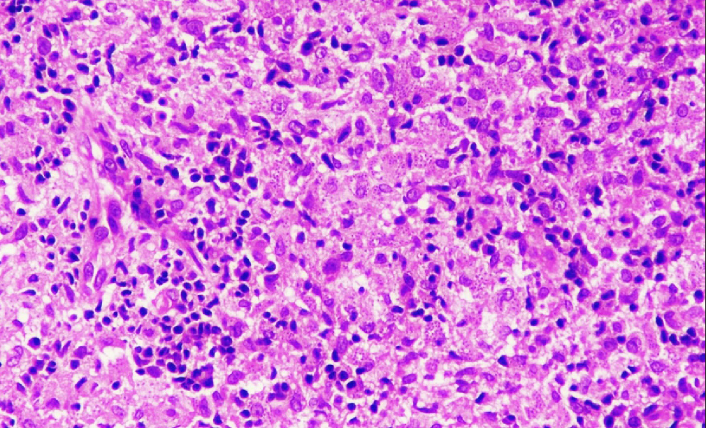
Entire dermis and subcutis show diffuse infiltrate of epithelioid histiocytes admixed with plasma cells and lymphocytes. Histiocytes are studded with small intra-cytoplasmic, basophilic inclusion bodies (H&E 100×)
Figure 2b
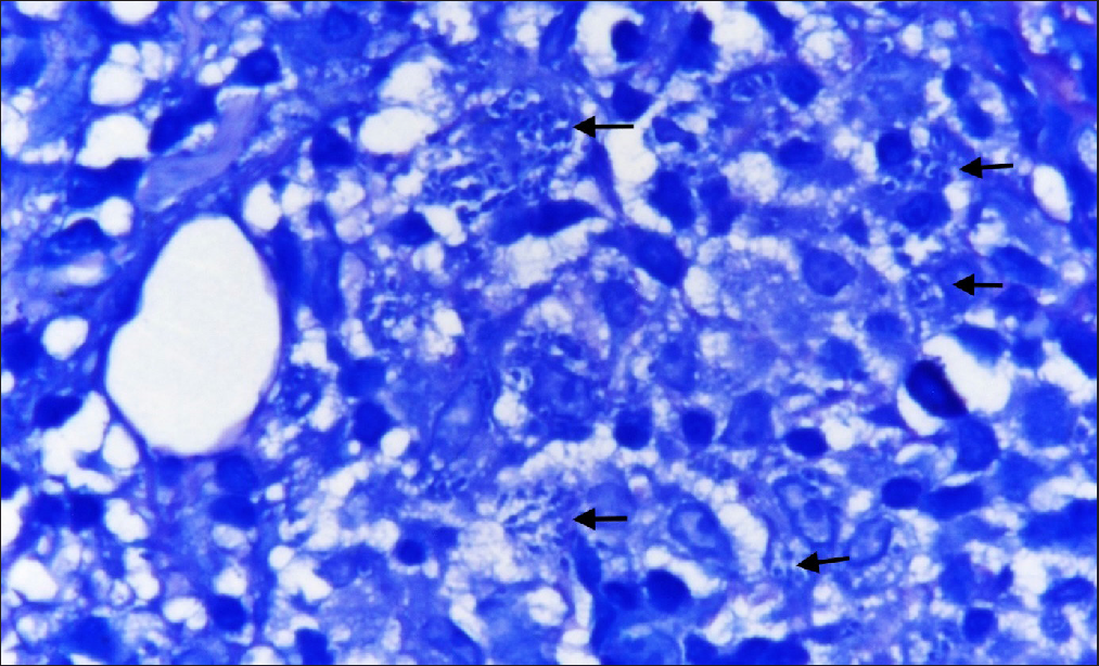
Arrows point towards intra-cellular inclusion bodies (Giemsa stain 400×)

Question

What is your Diagnosis?

Diagnosis

Localized Cutaneous Leishmaniasis

Discussion

Leishmaniasis is caused by protozoans of genus Leishmania and transmitted by sandflies. Leishmaniasis has mainly four clinical forms; cutaneous (diffuse or localized), mucosal, visceral and post-kala-azar dermal leishmaniasis. Other rare forms include leishmania recidivans and lupoid leishmaniasis. Cutaneous leishmaniasis is characterized by the development of single or multiple localized lesions on the exposed areas of skin after an incubation period of 3–8 weeks.1

Different species of Leishmania lead to different clinical features. Clinical forms of localized cutaneous leishmaniasis are papular, nodular, ulcerative plaque, infiltrative and impetigo-like.1 Face along with upper and lower extremities are the most commonly involved sites but ocular involvement is very rare. The eyelids, conjunctiva and inner parts of the eyes may be affected in patients having ocular involvement. Of these, eyelid leishmaniasis is very uncommon and is seen in only 2–5% of cases.2 There are only two published case reports of eyelid leishmaniasis from India.2,3

Regular movements of eyelids prevent sandfly bite in this area, leading to rarity of this disease on the eyelid. The differential diagnoses considered in this case were chalazion, lupus vulgaris, sarcoidosis, pseudo-lymphoma and basal cell carcinoma. Table 1 enumerates the clinical and histopathological differences among these entities.47 Lateral eyelid involvement is usually self-limiting and heals by scarring but medial eyelid involvement leads to contiguous spread to the conjunctiva, sclera and cornea with development of interstitial keratitis.3 Eyelid cutaneous leishmaniasis can lead to complications like palpebral scar, trichiasis, conjunctival scar, scleral perforation, corneal opacity, secondary glaucoma, epidermoid carcinoma and squamous cell carcinoma.1

Table 1 Differential diagnoses of eyelid leishmaniasis
Disease Clinical features Histopathological features
Chalazion4 Solitary, palpable, usually non-tender, non-fluctuant, non-erythematous nodule on the upper eyelid. Chronic granulomatous reaction with numerous lipid-filled, touton-type giant cells.
Lupus vulgaris5 Solitary well-defined plaque with central scarring and infiltration and scaling at margins. Tubercular granulomas with scanty or absent central caseation surrounded by epithelioid histiocytes and multinucleated giant cells that are present in the superficial dermis with prominent peripheral lymphocytes.
Sarcoidosis6 Solitary or multiple round or oval infiltrated plaques, brownish red in colour with little epidermal change. Dense, non-caseating naked granulomas composed of epithelioid histiocytes in the dermis which may extend into subcutaneous fat.
Pseudolymphoma7 Solitary or multiple smooth erythematous to bluish nodules typically located on the head, face or neck. Predominantly intradermal dense lymphocytic infiltrates (nodular or diffuse) consisting of small to medium-sized lymphocytes without significant nuclear atypia which may extend to the subcutis.
Basal cell carcinoma8 Usually present as a small papule which later forms a nodule which may be associated with ulcer and pigmentation. Classic palisading tumour. The various histological types of BCC include nodular (40–50%), infiltrative (15–25%), ulcerative, morphea-like, sclerosing, mixed, basosquamous and pigmented.

It is best to perform bed-side diagnostic test by taking a smear of the material from the lesion and staining it with Wright or Giemsa stain which may reveal the presence of Leishman-Donovan (LD) bodies. Histopathological examination of the lesion may also reveal LD bodies in early cases. Culture of the material in Novy-MacNeal-Nicolle medium may also be done for the confirmation of the diagnosis.3

Other modalities which can be used for diagnosis include leishmania intradermal skin test (Montenegro test), detection of anti-leishmania antibodies through agglutination or fluorescent antibody tests and polymerase chain reaction (PCR)-based methods including real-time PCR, DNA sequencing and through species-specific primers (DNA probes).9

Treatment modalities for cutaneous leishmaniasis include systemic or intralesional pentavalent antimony compounds, cryotherapy, topical paromomycin, local heat, excision, curettage, electrodessication, carbon dioxide laser, oral itraconazole, ketoconazole, miltefosine, etc.10 In the present case, she was started on oral itraconazole 200 mg twice daily. The lesion size reduced significantly after 2 months of treatment [Figure 3]. Itraconazole was continued further at the same dose for another 4 months which led to complete resolution of the lesion.

Figure 3
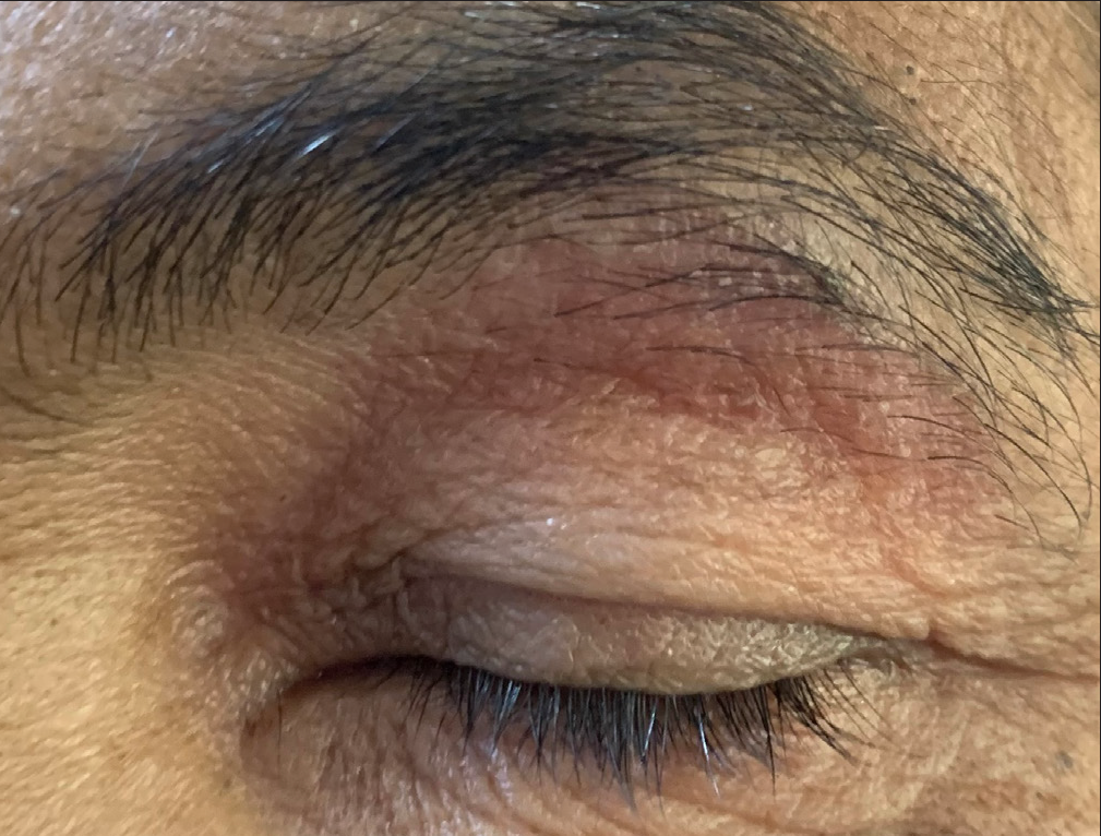
Decrease in erythema and size of the lesion after 2 months of treatment.

This case report highlights an unusual presentation of cutaneous leishmaniasis which clinicians need to be aware of.

Declaration of patient consent

The authors certify that they have obtained all appropriate patient consent.

Financial support and sponsorship

Nil.

Conflict of interest

There is no conflict of interest.

References

  1. , , , . Cutaneous leishmaniasis of the eyelids: a case series with molecular identification and literature review. Korean J Parasitol. 2016;54:787-92.
    [CrossRef] [PubMed] [PubMed Central] [Google Scholar]
  2. , , , , . Lid leishmaniasis: an atypical presentation of cutaneous leishmaniasis. Int J Health Sci Res. 2017;7:391-4.
    [Google Scholar]
  3. . Cutaneous leishmaniasis of the eyelid: an uncommon presentation of a common entity. Indian J Paediatr Dermatol. 2017;18:116-8.
    [Google Scholar]
  4. , , , , . Accuracy of the clinical diagnosis of chalazion. Eye (Lond). 2004;18:135-138.
    [CrossRef] [PubMed] [Google Scholar]
  5. , , . Lupus vulgaris: an atypical presentation. Indian J Dermatol Venereol Leprol. 2010;76:180-181.
    [CrossRef] [PubMed] [Google Scholar]
  6. , , , . Cutaneous sarcoidosis: clinical profile of 23 Indian patients. Indian J Dermatol Venereol Leprol. 2007;73:16-21.
    [CrossRef] [PubMed] [Google Scholar]
  7. , . Cutaneous pseudolymphoma-a review on the spectrum and a proposal for a new classification. J Cutan Pathol.. 2020;47:76-97.
    [CrossRef] [PubMed] [Google Scholar]
  8. , , , , . Basal cell carcinoma of eyelid. Arch Cytol Histopathol Res. 2019;4:102-4.
    [Google Scholar]
  9. , , , . Laboratory diagnosis of cutaneous and visceral leishmaniasis: current and future methods. Microorganisms. 2020;8:1632.
    [CrossRef] [PubMed] [PubMed Central] [Google Scholar]
  10. , , , , . Cutaneous leishmaniasis: successful treatment with itraconazole. Int J Dermatol. 2006;45:46-9.
    [CrossRef] [PubMed] [Google Scholar]

Fulltext Views
800

PDF downloads
286
View/Download PDF
Download Citations
BibTeX
RIS
Show Sections