Translate this page into:
Violaceous painful nodule of the leg in an Indian male patient
2 Department of Pathology, Fr. Muller Medical College, Mangalore, Karnataka, India
Correspondence Address:
M Ramesh Bhat
Department of Dermatology, Fr. Muller Medical College, Mangalore 2, Karnataka
India
| How to cite this article: Bhat M R, George AA, Pinto AC, Sukumar D, Lyngdoh RH. Violaceous painful nodule of the leg in an Indian male patient. Indian J Dermatol Venereol Leprol 2012;78:410 |
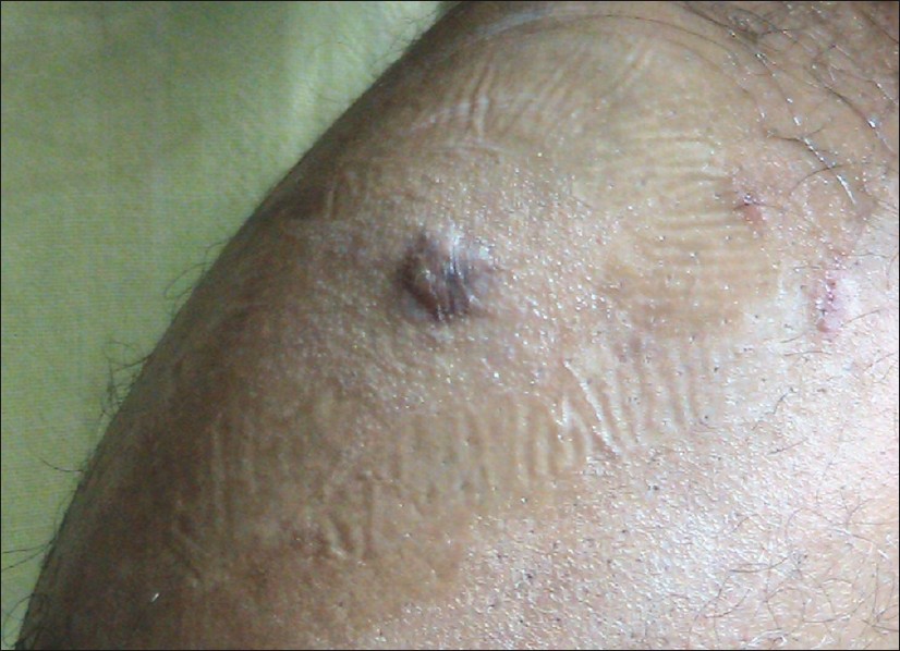
A 29-year-old man who was apparently normal presented with a 4-year history of painful lesion over the left leg below the knee. There was no history of preceding trauma. Patient complained of excruciating pain over the lesion on exposure to cold and even on application of very mild pressure. One year ago an attempt to excise the lesion was abandoned because of excruciating pain while giving local anesthesia. Cutaneous examination revealed an erythematous, tends cyanotic bluish nodule, 6 × 5 cms over left anteromedial aspect of calf about 9 cm below knee joint [Figure - 1]. Excision biopsy of the lesion was performed after infiltrating with local anesthetic. The specimen was sent for the histopathological examination.
 |
| Figure 1: Erythematous cyanotic nodule over left lower limb anteromedial aspect of the calf |
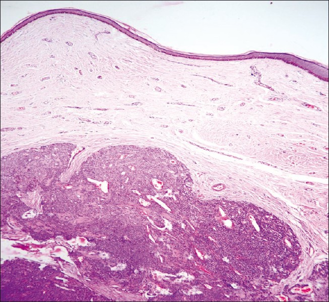
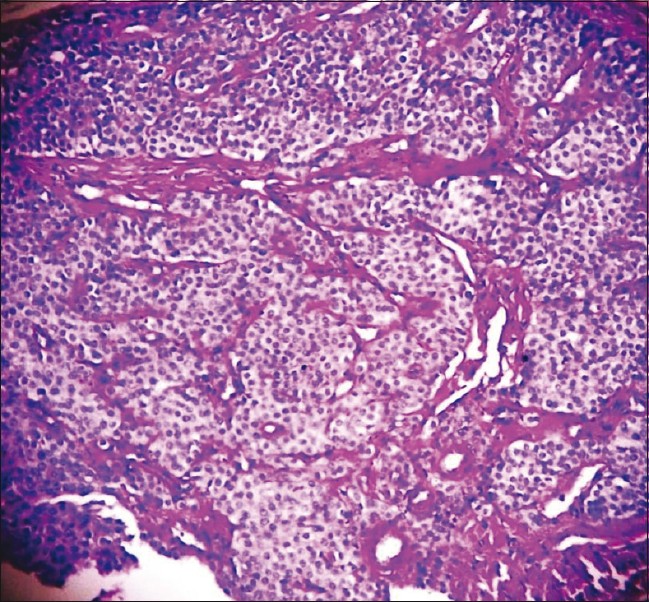
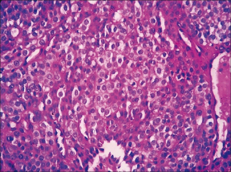
The histopathological examination showed well-defined and circumscribed solid tumor in the dermis extending to the subcutis [Figure - 2]. Tumor cells were arranged in solid sheets, nests which were surrounding scattered vascular channels ([Figure - 3], PAS counterstained with hematoxylin stain). The tumor was composed of uniformly round cells with well-defined cell borders with punched out darkly staining round to oval nuclei and clear to eosinophilic cytoplasm [Figure - 4]. There was presence of edematous stromal collagen. PAS and reticulin special stains were done for cell membrane and blood vessels.
 |
| Figure 2: Histopathology with H and E stain showing a well-defined tumor in the dermis with solid sheets of tumor cells which are surrounding vascular channels (×100) |
 |
| Figure 3: Histopathology with PAS counterstained with hematoxylin stain showing cells arranged in nests and surrounding vascular channels (×400) |
 |
| Figure 4: Histopathology with H and E stain showing solid sheets of uniform tumor cells Tumor cells are regular and rounded with eosinophilic cytoplasm and darkly staining round to oval nuclei (×400) |
What is Your Diagnosis ?
Answer: Glomus tumor
Discussion
Glomus tumors were first described clinically by Wood in 1812 as "painful subcutaneous tubercles". Glomus body is a neuromyoarterial body found within the reticular dermis that functions as a specialized form of arteriovenous anastamosis. The arterial end, or Sucquet - Hoyer canal is surrounded by uniform glomus cells. Glomus cells have properties similar to smooth muscle cells. Under normal conditions glomus bodies act to regulate blood flow to the skin. [1] Glomus bodies lie in the stratum reticularis of the dermis and are particularly numerous in the digits, palms and soles of the feet. [2]
Glomus tumors are neoplasms of normal glomus body. They classically present with a triad of symptoms- pain, pin-point tenderness with blunt palpation and hypersensitivity to cold. [1]
The various painful tumors of the skin are represented by the mnemonic BLEND AN EGG. [3] These tumors consist of a heterogenous group of proliferative tumors in the skin with pain as a single common factor. These tumors are (1) blue rubber bleb nevi, (2) leiomyoma cutis, (3) eccrinespiradenoma, (4) neuroma, (5) dermatofibroma, (6) angiolipoma, (7) neurilemmoma, (8) endometrioma, (9) glomus tumor, and (10) granular cell tumor. To this group tufted angioma also can be added which is also painful in nearly 30% of cases making the mnemonic BLEND TAN EGG. [4]
Glomus tumors are well-recognized cause of pain, particularly the subungual region. They have been thought to be much less common in extradigital locations. [1] Reviews of glomus tumors in the hand have shown a 2:1 female predominance and some studies have reported that as many as 75% of subungual lesions occur in females. [5] Extra digital glomus tumors appear to be found more commonly in males as in our case too. [1]
The histological examination reveals branching vascular channels separated by connective tissue stroma containing aggregates, nests and masses of specialized glomus cells that have electron microscopic features of smooth-muscle cells. Glomus tumors are most often of solid type, but vascular and myxoid types also occur. [2] Depending on the histology the tumor can be glomus tumor, glomangioma or glomangiomyoma. The stains for reticulin, nerve fibers, factor VIII, keratin, smooth muscle actin and neuroendocrine granules may be needed to clarify the diagnosis. Cure and pain alleviation with surgical excision is the rule. In our case, extradigital painful tumor in an Indian male patient was successfully excised without any recurrence.
| 1. |
Schiefer TK, Parker WL, Anakwenze OA, Amadio PC, Inwards CY, Spinner RJ. Extradigitalglomus tumors: A 20- year experience. Mayo ClinProc 2006;81:1337-44.
[Google Scholar]
|
| 2. |
Perks FJ, Beggs I, Lawson GM, Davie R. Juxtacorticalglomus tumor of the distal femur adjacent to the popliteal fossa. AmJ Radiol 2003;181:1590-2.
[Google Scholar]
|
| 3. |
Kaleeswaran AV, Janaki VR, Sentamilselvi G, Mohan CK. Eccrinespiradenoma. Indian J Dermatol Venereol Leprol 2002;68:236-7.
[Google Scholar]
|
| 4. |
Kamath GH, Bhat RM, Kumar S. Tufted angioma. Int J Dermatol 2005;44:1045-7.
[Google Scholar]
|
| 5. |
Maxwel GP, Curtis RM, Wilgis EF. Multiple digital glomus tumors. J Hand SurgAm 1979;4:363-7.
[Google Scholar]
|
Fulltext Views
3,837
PDF downloads
3,624


