Translate this page into:
Zoster ophthalmicus with dissemination in a six year old immunocompetent child
Correspondence Address:
Subhash Kashyap
Department of Dermatology, Indira Gandhi Medical College, Shimla - 171 001, Himachal Pradesh
India
| How to cite this article: Kashyap S, Shanker V. Zoster ophthalmicus with dissemination in a six year old immunocompetent child. Indian J Dermatol Venereol Leprol 2014;80:382 |
Sir,
Herpes zoster ophthalmicus is defined as herpes zoster involving the ophthalmic division of the fifth cranial nerve. Following primary varicella (chickenpox) infection, the virus remains dormant in the dorsal root or other sensory ganglia. Reactivation usually occurs due to a decline in the specific cell-mediated immunity against varicella-zoster virus (VZV) with aging, immunosuppression or both. [1] Though it is reported to occur as early as the first week of life, about 66% of patients with herpes zoster are older than 50 years, and only 5% are younger than 15. [2] The condition in childhood differs from that in adults in that it is milder, without a prodrome, and is generally not associated with postherpetic neuralgia. [2] We report an unusual case of herpes zoster ophthalmicus with dissemination in a six-year-old immunocompetent child, whose mother had chickenpox when she was pregnant with him.
A six-year-old boy presented to us with vesicles and severe crusting predominantly involving the left side of his upper face and scalp for about 20 days. Fluid-filled lesions had appeared abruptly on the left eyelids and spread to the ipsilateral forehead and scalp within a day. A few scattered lesions had then appeared on his right cheek as well as on the shoulder and trunk in the next two days. The child was not on any medication and there was no history suggestive of imunosupression or malignancy. There was also no history suggestive of a previous episode of chickenpox and he was not immunized against varicella. The patient′s mother gave a history of having had chickenpox while the child was six months in utero, the antenatal history being otherwise unremarkable.
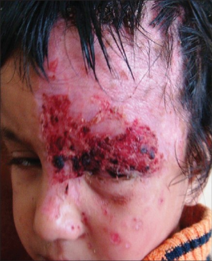
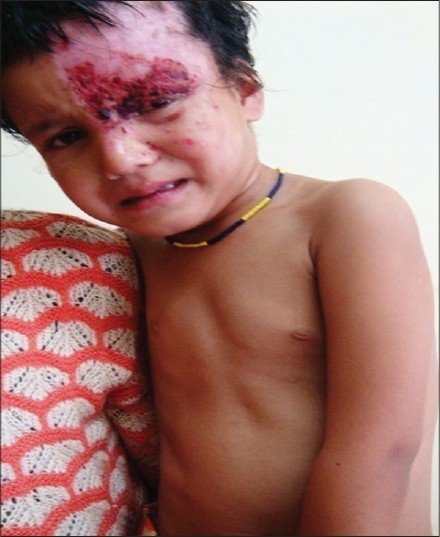
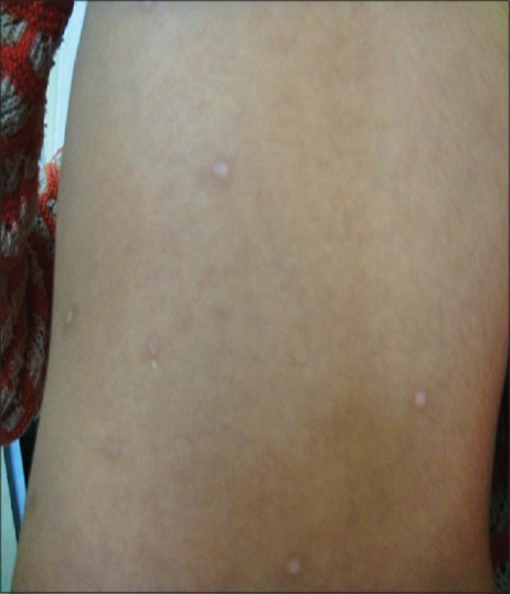
On examination, there were multiple vesicles and hemorrhagic crusts on the left frontal area coalescing centrally into a well-demarcated lesion and extending to involve the left cheek, eyelids, and temporo-occipital areas [Figure - 1]. Similar discrete lesions (>20 in number) and healed hypopigmented varioliform scars were present over the right cheek, shoulders and trunk [Figure - 2] and [Figure - 3]. The left eye was congested and there was a sealed perforation at 4 o′clock with the iris incarcerated in the wound. Clinically, there was no suggestion of malnutrition.
 |
| Figure 1: Severe crusting involving the area supplied by the ophthalmic nerve. Similar lesions are also seen on the nose and both cheeks |
 |
| Figure 2: Discrete healed hypopigmented lesions seen over the trunk and left shoulder. Lesions other than those of HZO were more than 20 in number |
 |
| Figure 3: A closer view showing healed lesions on the left deltoid region |
Routine investigations including a complete hemogram and chest X-rays were normal. A Tzanck smear from the vesicular lesions was positive for multinucleated giant cells. HIV serology was negative in both mother and child but the parents could not afford to have serum immunoglobulin levels tested. Based on the clinical features, a diagnosis of zoster ophthalmicus with dissemination was made and the child was managed with oral acyclovir and supportive treatment. The skin lesions recovered within a week with residual scarring and alopecia. Ophthalmologic advice was sought for management of the eye lesions which eventually healed with residual visual impairment due to corneal scarring.
The timing of primary VZV infection in pregnancy determines the risk of fetal involvement with infection between 8 and 20 weeks of gestation leading to the congenital varicella syndrome in 12% of infants. These infants are born with cicatricial lesions, cutaneous defects and hypopigmentation in addition to multi-organ involvement. [3] Intrauterine growth retardation and preterm birth may also occur. In the remaining cases and in infections at a later stage of pregnancy, the fetus is either not infected or suffers a subclinical infection with persistence of immunoglobulin G (IgG) at 1-2 years of age. The immunological status at the time of acquiring the primary infection is the most important determinant in subsequent viral reactivation. The low levels of lymphocytes, natural killer (NK) cells, cytokines and virus specific immunoglobulins seen in utero and in infancy may result in an inability to maintain the latency of VZV leading to the early appearance of zoster in children. [4]
The incidence of herpes zoster in otherwise healthy children is rising of late. [3] This may be due to an increase in primary varicella infection in utero, in infancy or due to vaccination with the live attenuated virus. The diagnosis of herpes zoster is usually made clinically but should be confirmed by Tzanck smear, direct fluorescent antibody tests, high or rising titers of antibodies against VZV, or by culture studies. Herpes zoster in childhood should be differentiated from zosteriform herpes simplex, bullous impetigo and bullous insect bite reactions. [4] Herpes zoster ophthalmicus in childhood with HIV can be a systemic disease and may be associated with tuberculosis and syphilis. [5] Though systemic disease and prodromal symptoms were absent in our case, disseminated skin involvement and serious local complications did occur. This case is reported to highlight the fact that severe herpes zoster ophthalmicus can occur even in immunocompetent children without a history of varicella, as primary infection can occur in utero.
| 1. |
Sanjay S, Huang P, Lavanya R. Herpes zoster ophthalmicus. Curr Treat Options Neurol 2011;13:79-91.
[Google Scholar]
|
| 2. |
Aikenhead KJ, Johnson TL Jr. Herpes zoster in a 6-month-old infant with 13-year follow-up: A retrospective case report. J Chiropr Med 2011;10:306-9.
[Google Scholar]
|
| 3. |
Sanchez MA, Bello-Munoz JC, Cebrecos I, Sanz TH, Martinez JS, Moratonas EC, et al. The prevalence of congenital varicella syndrome after a maternal infection, but before 20 weeks of pregnancy: A prospective cohort study. J Matern Fetal Neonatal Med 2011;24:341-7.
[Google Scholar]
|
| 4. |
Bhushan P, Sardana K, Mahajan S. Dermatomal vesicular eruption in an asymptomatic infant. Dermatol Online J 2005;11:26.
[Google Scholar]
|
| 5. |
Gupta N, Sachdev R, Sinha R, Titiyal JS, Tandon R. Herpes zoster ophthalmicus: Disease spectrum in young adults. Middle East Afr J Ophthalmol 2011;18:178-82.
[Google Scholar]
|
Fulltext Views
2,941
PDF downloads
2,459


