Generic selectors
Exact matches only
Search in title
Search in content
Post Type Selectors
Search in posts
Search in pages
Filter by Categories
15th National Conference of the IAOMFP, Chennai, 2006
Abstract
Abstracts from current literature
Acne in India: Guidelines for management - IAA Consensus Document
Addendum
Announcement
Art & Psychiatry
Article
Articles
Association Activities
Association Notes
Award Article
Book Review
Brief Report
Case Analysis
Case Letter
Case Letters
Case Notes
Case Report
Case Reports
Clinical and Laboratory Investigations
Clinical Article
Clinical Studies
Clinical Study
Commentary
Conference Oration
Conference Summary
Continuing Medical Education
Correspondence
Corrigendum
Cosmetic Dermatology
Cosmetology
Current Best Evidence
Current Issue
Current View
Derma Quest
Dermato Surgery
Dermatopathology
Dermatosurgery Specials
Dispensing Pearl
Do you know?
Drug Dialogues
e-IJDVL
Editor Speaks
Editorial
Editorial Remarks
Editorial Report
Editorial Report - 2007
Editorial report for 2004-2005
Errata
Erratum
Focus
Fourth All India Conference Programme
From Our Book Shelf
From the Desk of Chief Editor
General
Get Set for Net
Get set for the net
Guest Article
Guest Editorial
History
How I Manage?
IADVL Announcement
IADVL Announcements
IJDVL Awards
IJDVL AWARDS 2015
IJDVL Awards 2018
IJDVL Awards 2019
IJDVL Awards 2020
IJDVL International Awards 2018
Images in Clinical Practice
Images in Dermatology
In Memorium
Inaugural Address
Index
Knowledge From World Contemporaries
Leprosy Section
Letter in Response to Previous Publication
Letter to Editor
Letter to the Editor
Letter to the Editor - Case Letter
Letter to the Editor - Letter in Response to Published Article
LETTER TO THE EDITOR - LETTERS IN RESPONSE TO PUBLISHED ARTICLES
Letter to the Editor - Observation Letter
Letter to the Editor - Study Letter
Letter to the Editor - Therapy Letter
Letter to the Editor: Articles in Response to Previously Published Articles
Letters in Response to Previous Publication
Letters to the Editor
Letters to the Editor - Letter in Response to Previously Published Articles
Letters to the Editor: Case Letters
Letters to the Editor: Letters in Response to Previously Published Articles
Media and news
Medicolegal Window
Messages
Miscellaneous Letter
Musings
Net Case
Net case report
Net Image
Net Images
Net Letter
Net Quiz
Net Study
New Preparations
News
News & Views
Obituary
Observation Letter
Observation Letters
Oration
Original Article
ORIGINAL CONTRIBUTION
Original Contributions
Pattern of Skin Diseases
Pearls
Pediatric Dermatology
Pediatric Rounds
Perspective
Presedential Address
Presidential Address
Presidents Remarks
Quiz
Recommendations
Regret
Report
Report of chief editor
Report of Hon : Treasurer IADVL
Report of Hon. General Secretary IADVL
Research Methdology
Research Methodology
Resident page
Resident's Page
Resident’s Page
Residents' Corner
Residents' Corner
Residents' Page
Retraction
Review
Review Article
Review Articles
Reviewers 2022
Revision Corner
Self Assessment Programme
SEMINAR
Seminar: Chronic Arsenicosis in India
Seminar: HIV Infection
Short Communication
Short Communications
Short Report
Snippets
Special Article
Specialty Interface
Studies
Study Letter
Study Letters
Supplement-Photoprotection
Supplement-Psoriasis
Symposium - Contact Dermatitis
Symposium - Lasers
Symposium - Pediatric Dermatoses
Symposium - Psoriasis
Symposium - Vesicobullous Disorders
SYMPOSIUM - VITILIGO
Symposium Aesthetic Surgery
Symposium Dermatopathology
Symposium-Hair Disorders
Symposium-Nails Part I
Symposium-Nails-Part II
Systematic Review and Meta-Analysis
Systematic Reviews and Meta-analyses
Systematic Reviews and Meta-analysis
Tables
Technology
Therapeutic Guideline-IADVL
Therapeutic Guidelines
Therapeutic Guidelines - IADVL
Therapeutics
Therapy
Therapy Letter
Therapy Letters
View Point
Viewpoint
What’s new in Dermatology
View/Download PDF

Translate this page into:

Resident’s page
90 (
6
); 838-841
doi:
10.25259/IJDVL_1308_2023
pmid:
38841957

Ultraviolet reflectance dermoscopy

Department of Dermatology, Sree Narayana Institute of Medical Sciences, Kochi, Kerala, India
Department of Dermatology, Medical Trust Hospital, Cochin, Kerala, India
Department of Dermatology, Amanza Skin Clinic, Perinthalmanna, Kerala, India

Corresponding author: Dr. Feroze Kaliyadan, Department of Dermatology, Sree Narayana Institute of Medical Sciences, Kochi, Kerala, India. ferozkal@gmail.com

Licence
This is an open-access article distributed under the terms of the Creative Commons Attribution-Non Commercial-Share Alike 4.0 License, which allows others to remix, transform, and build upon the work non-commercially, as long as the author is credited and the new creations are licensed under the identical terms.

How to cite this article: Kaliyadan F, Jayasree P, Ashique KT. Ultraviolet reflectance dermoscopy. Indian J Dermatol Venereol Leprol. 2024;90:838-41. doi: 10.25259/IJDVL_1308_2023

Introduction

Ultraviolet (UV) light has been an integral part of the dermatological examination, primarily in the form of the Wood’s lamp, which uses ‘black light’ having a peak output wavelength of 365 nm and aids in the diagnosis of conditions like pigmentary disorders (vitiligo, melasma, naevus depigmentosus) and superficial (fungal and corynebacterial) infections. Some other indications for which the Wood’s lamp can be used include – evaluation of Cutibacterium acnes, which can produce an orange-red colour in pilosebaceous follicles, pseudomonas infections which can produce a green fluorescence, and also porphyrias.1 The Wood’s lamp can also be used in the evaluation of premature canities and compliance regarding the use of sunscreens.2,3

UV dermoscopy

UV reflectance dermoscopy (UVRD) is a relatively new innovation, combining dermoscopy with UV light evaluation. Like the Wood’s lamp, UV dermoscopy can be useful in pigmentary disorders such as vitiligo and melasma. Lesional margins, perifollicular pigmentation, and depigmentation can be better appreciated under UV dermoscopy as compared to normal dermoscopy.2 Dermoscopy with near-UV light can highlight demarcation of the distribution of melanin in cutaneous melanoma.4

Sebaceous gland openings can show fluorescence under UVRD – ranging from bright blue/green to yellow, orange, or red. This could possibly be useful in the diagnosis and follow-up of acne vulgaris [Figure 1]. The same principle has been used to differentiate Fordyce’s spots from its mimickers. On normal polarised dermoscopy, Fordyce’s spots present as whitish-yellow clods, clustered together. Under UVRD, the fluorescence at the tip of the sebaceous opening can be seen over the less prominent, ill-defined yellow-green clods.5
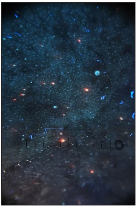
Orange fluorescence due to porphyrins produced by cutibacterium acnes (Dermlite™ DL5 UV mode).
Figure 1:
Orange fluorescence due to porphyrins produced by cutibacterium acnes (Dermlite™ DL5 UV mode).

UVRD has been shown to be useful in the diagnosis of scabies too. UVRD of scabietic burrows highlights the blue fluorescence of the burrow and can also visualise a bright green fluorescence from the female mite’s body.6

UVRD showing prominent yellowish-green fluorescence has been reported in trichomycosis axillaris.7 Pink glow on UV dermoscopy has been identified as a helpful sign for the diagnosis of subungual glomus tumour.8

Other applications of UVRD

Some other indications in which we found UVRD useful include:

  • Evaluation of premature canities - like the Wood’s lamp examination, UVRD can highlight the grey hairs better. Similarly, it can help in the evaluation of fine regrowing hairs in alopecia areata (AA). Fine, regrowing hairs in AA can be devoid of pigment. UVRD picks up these hairs better as compared to standard dermoscopy [Figures 2a and 2b].

  • Molluscum contagiosum – central umbilicated area shows bright white fluorescence [Figures 3a and 3b].

  • Scaling tends to be generally more prominently seen under UV dermoscopy. For example, in pityriasis versicolor – the fines scales are visible more clearly using UVRD and can be enhanced by grattage test [Figures 4a and 4b].

  • The UV blink sign - The dermoscopic ‘blink sign’ describes the blinking appearance of certain dermoscopic patterns when the device is toggled between polarised and non-polarised modes. For example, the comedo-like openings in seborrheic keratosis are visualised more clearly on non-polarised dermoscopy, as compared to polarised dermoscopy, and hence these structures appear to blink when the dermoscope is toggled between polarised and non-polarised modes.9 The comedo-like openings and milia-like cysts seen in seborrheic keratosis tend to be clearer under UV and these structures show a blinking effect when the dermoscope is toggled between UV and non-UV modes. We would like to label this as the ‘dermoscopic UV blink sign’ (personal observation) [Figures 5a and 5b].

  • The pigmentary patterns in conditions like vitiligo tend to be generally clearer under UV dermoscopy [Figures 6a and 6b].
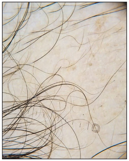
Alopecia areata showing regrowing hairs (Dermlite ™ DL5 polarised mode).
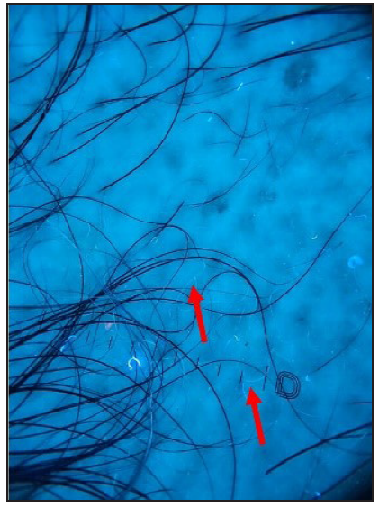
Figure 2a:
Alopecia areata showing regrowing hairs (Dermlite ™ DL5 polarised mode).
White hairs are seen more clearly under UV dermoscopy (red arrows) (Dermlite ™ DL5 UV mode).
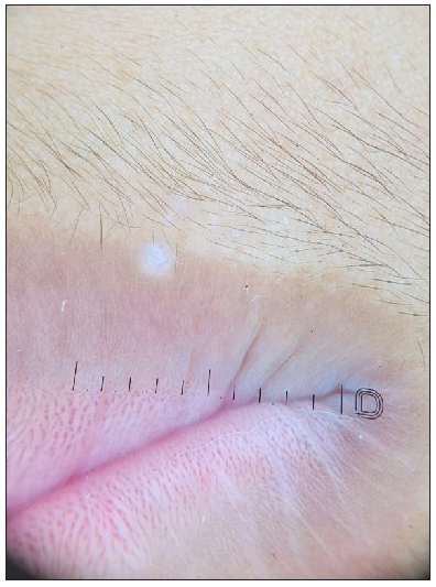
Figure 2b:
White hairs are seen more clearly under UV dermoscopy (red arrows) (Dermlite ™ DL5 UV mode).
Dermoscopy of molluscum contagiosum (Dermlite ™ DL5 polarised mode).
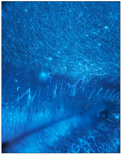
Figure 3a:
Dermoscopy of molluscum contagiosum (Dermlite ™ DL5 polarised mode).
Dermoscopy of molluscum contagiosum – central umbilicated area shows bright white fluorescence (Dermlite ™ DL5 UV mode).
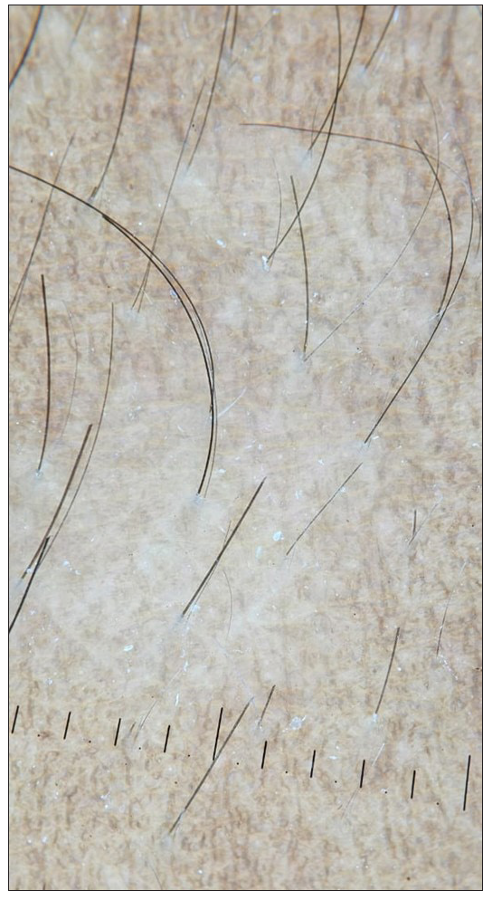
Figure 3b:
Dermoscopy of molluscum contagiosum – central umbilicated area shows bright white fluorescence (Dermlite ™ DL5 UV mode).
Dermoscopy of hypopigmented pityriasis versicolor (Dermlite™ DL5 polarised mode).
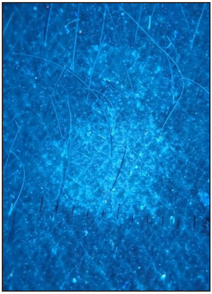
Figure 4a:
Dermoscopy of hypopigmented pityriasis versicolor (Dermlite™ DL5 polarised mode).
Dermoscopy of hypopigmented pityriasis versicolor – fines scales are visible more clearly (Dermlite ™ DL5 UV mode).
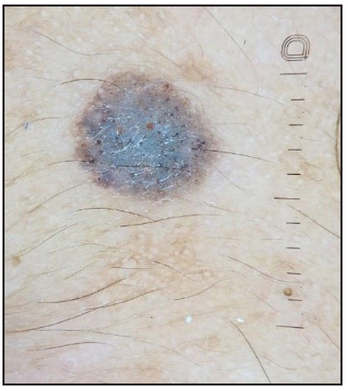
Figure 4b:
Dermoscopy of hypopigmented pityriasis versicolor – fines scales are visible more clearly (Dermlite ™ DL5 UV mode).
The UV dermoscopic blink sign in seborrheic keratosis (Dermlite ™ DL5 polarised mode).
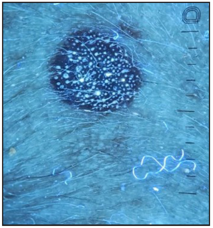
Figure 5a:
The UV dermoscopic blink sign in seborrheic keratosis (Dermlite ™ DL5 polarised mode).
The UV dermoscopic blink sign in seborrheic keratosis (Dermlite ™ DL5 UV mode).
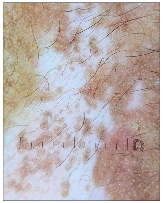
Figure 5b:
The UV dermoscopic blink sign in seborrheic keratosis (Dermlite ™ DL5 UV mode).
Dermoscopy of vitiligo (Dermlite™ DL5 polarised mode).
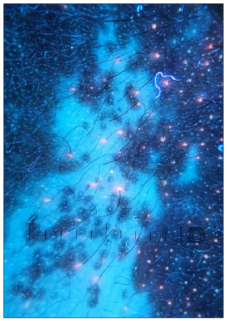
Figure 6a:
Dermoscopy of vitiligo (Dermlite™ DL5 polarised mode).
Dermoscopy of vitiligo – white glow seen more clearly (Dermlite ™ DL5 UV mode).
Figure 6b:
Dermoscopy of vitiligo – white glow seen more clearly (Dermlite ™ DL5 UV mode).

Conclusion

The combination of dermoscopy with UV light is a relatively recent introduction and can be useful in diagnostic dermatology. Most dermoscopes that incorporate UV light use a non-polarised form of UV light and hence it is basically like an examination under magnification using UV light. However, using a contact fluid along with the UV light mode will convert this into true UV dermoscopy

Declaration of patient consent

The authors certify that they have obtained all appropriate patient consent.

Financial support and sponsorship

Nil.

Conflicts of interest

There are no conflicts of interest.

Use of artificial intelligence (AI)-assisted technology for manuscript preparation

The authors confirm that there was no use of artificial intelligence (AI)-assisted technology for assisting in the writing or editing of the manuscript and no images were manipulated using AI.

References

  1. , . Revealing the unseen: A review of wood’s lamp in dermatology. J Clin Aesthet Dermatol. 2022;15:25-30.
    [PubMed] [PubMed Central] [Google Scholar]
  2. , , , , , . Novel ultraviolet-dermoscopy: Early diagnosis and activity evaluation of vitiligo. Skin Res Technol. 2023;29:e13249.
    [CrossRef] [PubMed] [PubMed Central] [Google Scholar]
  3. , , . Looking for a grey needle in a ‘hair’ stack! Using a wood’s lamp for evaluating canities. Indian J Dermatol Venereol Leprol. 2023;89:636-7.
    [CrossRef] [PubMed] [Google Scholar]
  4. , , , , . Dermoscopy with near-ultraviolet light highlights the demarcation of melanin distribution in cutaneous melanoma. J Am Acad Dermatol. 2021;84:e23-e24.
    [CrossRef] [PubMed] [Google Scholar]
  5. , , , , , . Differentiating fordyce spots from their common simulators using ultraviolet-induced fluorescence dermatoscopy-retrospective study. Diagnostics (Basel). 2023;13:985.
    [CrossRef] [PubMed] [PubMed Central] [Google Scholar]
  6. , . Scabies mite is bright green under uv dermatoscopy. Dermatol Pract Concept. 2023;13:e2023135.
    [CrossRef] [PubMed] [PubMed Central] [Google Scholar]
  7. , , , , , . Ultraviolet reflectance dermatoscopy of trichobacteriosis axillaris reveals peripilar yellow-green luminescent concretions. Dermatol Pract Concept. 2023;13:e2023169.
    [CrossRef] [PubMed] [PubMed Central] [Google Scholar]
  8. , , . “Pink glow”: A new sign for the diagnosis of glomus tumor on ultraviolet light dermoscopy. Indian Dermatol Online J. 2015;6:S21-3.
    [CrossRef] [PubMed] [Google Scholar]
  9. , , . The “blink sign” in dermoscopy. Arch Dermatol. 2011;147:520.
    [CrossRef] [PubMed] [Google Scholar]
Show Sections