Translate this page into:
Gentian violet: Revisited
2 Department of ENT and HNS, All India Institute of Medical Sciences, Raipur, Chhattisgarh, India
Correspondence Address:
Neel Prabha
Department of Dermatology, All India Institute of Medical Sciences, GE Rd., Tatibandh, Raipur, Chhattisgarh
India
| How to cite this article: Prabha N, Arora RD, Ganguly S, Chhabra N. Gentian violet: Revisited. Indian J Dermatol Venereol Leprol 2020;86:600-603 |
Gentian violet, also known as crystal violet or methyl violet, is a triphenylmethane dye. It is commonly used as a stain in laboratory. Gentian violet was first synthesized in 1861 by French chemist Charles Lauth.[1] Hans Gram in 1884 noticed its irreversible fixation by Gram positive bacteria.[2] In 1891, it was introduced as an antiseptic by Stilling. Its antibacterial action against Gram positive bacteria was noticed by Churchman in 1912.[3] Since then, it was used in a variety of diseases because of its antibacterial, antifungal, anti-helminthic and anti-trypanosomal properties. Following discovery of penicillin and sulfa drugs, its use has declined.
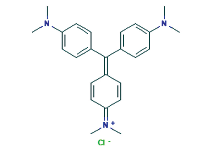
Gentian violet is a symmetric compound with 6 methyl groups. Chemical formula of gentian violet is [4-[bis[4-(dimethylamino) phenyl] methylidene] cyclohexa-2,5-dien-1-ylidene]-dimethylazanium chloride [Figure - 1].[4]
 |
| Figure 1: Chemical structure of gentian violet |
For topical use gentian violet is available commercially as 0.5% and 1% aqueous or alcohol solution. Gentian violet and methylene blue antibacterial dressings in polyvinyl alcohol foam and polyurethane foam are also available. Gentian violet as 0.5% aqueous solution is licensed for topical application on unbroken skin but is not recommended for application on mucous membranes or open wounds.[5] This review discusses the role of gentian violet in dermatology.
Indications of Gentian Violet in Dermatology
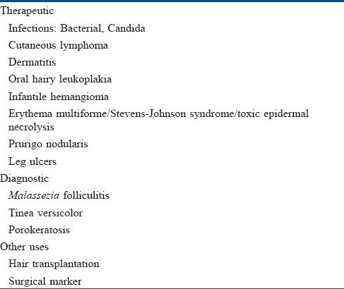
Indications of gentian violet in dermatology are given in [Table - 1].

Infections
Gentian violet is effective against Streptococcus, Staphylococcus species, methicillin-resistant Staphylococcus aureus and moderately effective against gram negative bacteria.[6],[7],[8] Gentian violet also inhibits the growth of Pseudomonas and disrupts Pseudomonas biofilms in vitro.[6],[9],[10]
Multiple hypotheses for its antibacterial action are given. The bacteriostatic effect is due to the unfavorable oxidation-reduction potential poised by gentian violet and the inhibition of reduced nicotinamide adenine dinucleotides (NADPH) oxidases by it.[11],[12] It penetrates the bacterial cell wall and forms a covalent adduct with thioredoxin reductase 2 which is essential for cellular activity, ultimately leading to cell death.[13] It reduces vascular leak by reducing the levels of angiopoietin-2 expression and allows improved antibiotic delivery.[14]
Gentian violet is also effective against Candida.[15],[16],[17]
Lymphoma
Gentian violet has been tried in lymphoma patients. Regression of primary cutaneous diffuse B cell lymphoma after a single injection of intralesional gentian violet has been reported in a patient who was unfit for conventional therapy.[18] In another case report, partial clinical response of recalcitrant, localized, stage 1B mycosis fungoides has been reported.[19] The mechanism for antitumor activity of gentian violet is not fully known. It inhibits reduced NADPH oxidase, an enzyme that converts molecular oxygen to superoxide and hydrogen peroxide. By inhibiting this enzyme, gentian violet decreases local reactive oxygen species, which in turn might inhibit certain tumor suppressor genes like PTEN, Ik-B and p53.[13] Gentian violet also inhibits NF-kB, possibly through NADPH oxidase inhibition, which has been implicated in proliferation of cutaneous T-cell lymphoma tumor cells.[19] Wu and Wood found that gentian violet enhances Fas and TRAIL pathway dependent apoptosis of cutaneous T-cell lymphoma cell lines and tumor cells in Sezary syndrome.[20]
Dermatitis
Gentian violet is also helpful in atopic eczema. In atopic eczema, skin colonization with Staphylococcus aureus and high levels of proinflammatory angiopoietin-2 plays a possible role in the pathophysiology of the disease. Gentian violet by decreasing bacterial colonization and angiopoietin-2 levels reduces the severity of the eczema.[21]
Gentian violet may be used in treatment of irritant dermatitis. Gloor et al. noted anti-irritative effect of 0.5% gentian violet.[22] They found that 0.5% gentian violet reduces skin damage in irritative dermatitis. It also provides immediate pain relief in acute painful eczematous lesions.[23]
Oral hairy leukoplakia
Gentian violet application can be used to treat oral hairy leukoplakia.[24] The role of gentian violet in oral hairy leukoplakia is based on the evidence that Epstein bar virus oncogenes induce the generation of reactive oxygen species, and gentian violet is a potent inhibitor of reduced NADPH oxidase which generates these reactive oxygen.[24]
Hair transplantation
Gentian violet can be used for visualizing white hair during the punching procedure and graft preparation in follicular unit extraction for white-haired patients.[25] The follicular units of white hair are too white or transparent to be distinguished from the surrounding soft tissue. However, after 1% gentian violet dyeing of the scalp skin, the soft tissue is also dyed, allowing for a clear view while trimming and loading into the implanter.
Infantile hemangioma
Gentian violet may accelerate healing of ulcerated infantile hemangioma.[26] Ulceration in infantile hemangiomas is due to an imbalance between angiopoietin-2 and vascular endothelial growth factor. Angiopoietin-2 promotes angiogenesis in the presence of vascular endothelial growth factor. Gentian violet decreases the production of angiopoietin-2 and promotes ulcer healing.[26]
Erythema multiforme/Stevens-Johnson syndrome/toxic epidermal necrolysis
Topical gentian violet stabilizes and halts the progression of new cutaneous bullae of erythema multiforme.[27] Angiopoietin-2 induced vascular leak is involved in the pathogenesis of erythema multiforme. Nitric oxide and toxic peroxynitrite are involved in keratinocyte necrosis occuring in this condition. Gentian violet down-regulates the production of angiopoietin-2 and peroxynitrite.[27] Gupta et al. 28 recommended the use of gentian violet paint in dilution for treating denuded areas in SJS/TEN.
Prurigo nodularis
Gentian violet may be effective in treating prurigo nodularis. Prurigo nodularis is accompanied by STAT6 and STAT3 activation. Gentian violet may be effective in this condition by targeting STAT 6 signal.[29]
Leg ulcers
Antibacterial foam dressing consisting of polyvinyl alcohol bound to gentian violet and methylene blue (GV/MB PVA) can be a suitable option for chronic leg ulcers, especially diabetic foot ulcers.[30]
Polyurethane foam bound gentian violet and methylene blue (GV/MB PU) dressings are also available commercially. Gentian violet-methylene blue in polyvinyl alcohol foam requires saline hydration, whereas that in polyurethane foam dressing, do not. Gentian violet-methylene blue in polyurethane foam dressings are best suited for moist wounds that do not require additional hydration.[30] These dressings absorb and trap bacterial debris away from the wound, aid in autolytic debridement and thus promote re-epithelialization by flattening the wound edges.[31]
Gram staining
Gentian violet is used as a stain in Gram staining method. This staining method helps in differentiating Gram positive and Gram negative bacteria. Gram staining is also helpful in the diagnosis of Malasezzia folliculitis.[32]
Other Uses
In vivo gram staining is a bedside diagnostic test for tinea versicolor.[33] In this test, when gentian violet is applied to the site of tinea versicolor, a dramatic accentuation of the infected areas compared with unaffected skin occurs due of retention of the dye by the fungus. This test differentiates tinea versicolor from other conditions like atopic dermatitis, vitiligo and pityriasis rosea as these conditions do not retain gentian violet.[33] Ink test can be performed for diagnosing porokeratosis. Circumferential furrow of the lesion can be delineated by applying gentian violet to the lesions of porokeratosis.[34]
Gentian violet solution is also used in surgery to mark the surgical sites. Chen et al.[35] described a sterile, effective and economical intraoperative skin marking method by using gentian violet. Four drops of gentian violet was dispensed into microcentrifuge tube, which was then autoclaved after capping. Toothpick was used as the writing instrument. The advantage of this method was that unlike commercially available skin markers, skin moisture did not cause the writing implement to become ineffective.
Side Effects
Common side effect is staining by gentian violet. There are reports of irritant contact dermatitis with gentian violet 3% preparation.[36],[37] The predisposing factors mentioned in these reports were application on intertriginous area and prolonged contact. Pasricha et al.[38] and Bajaj et al.[39] found contact sensitivity to gentian violet. Oral ulceration was reported with its application in oral candidiasis.[40],[41] A woman developed severe hemorrhagic cystitis due to accidental injection of gentian violet through the urethra.[42] A patient developed superficial necrosis of the glans penis after topical treatment with 1% gentian violet.[43] Accidental instillation of gentian violet in eye can lead to keratoconjunctivitis.[44]
Some consider gentian violet as a potential carcinogen. Rosenkranz et al.[45] found that gentian violet interacted with the DNA of living cells. They opined that it reacted with the cellular DNA which might induce detrimental changes. Animal studies performed on mice and rats found that increased rate of hepatocellular and thyroid cancer was seen when gentian violet was fed in large doses for long duration.[46],[47] However, no reports of cancer in human associated with topical gentian violet use have been found in the recent literature.
Conclusion
Gentian violet, in the present era of emerging resistance, is a cheaper and safer alternative to topical antibiotics. As it has anti-angiogenic, anti-tumor and anti-inflammatory properties, it can be used as an adjuvant in other dermatoses. To prevent side effects, it should be applied unoccluded, for a short contact-time and should be kept away from the eyes during application.
Financial support and sponsorship
Nil.
Conflicts of interest
There are no conflicts of interest.
| 1. |
Lauth C. On the new aniline dye, Violet de Paris. Laboratory 1867;1:138-9.
[Google Scholar]
|
| 2. |
Balabanova M, Popova L, Tchipeva R. Dyes in dermatology. Clin Dermatol 2003;21:2-6.
[Google Scholar]
|
| 3. |
Churchman JW. The selective bactericidal action of gentian violet. J Exp Med 1912;16:221-47.
[Google Scholar]
|
| 4. |
Available from: https://pubchem.ncbi.nlm.nih.gov/compound/Gentian-violet. [Last accessed on 2020 Jan 04].
[Google Scholar]
|
| 5. |
Berth-Jones J. Principles of topical therapy. In: Griffiths C, Barker J, Bleiker T, Chalmers R, Creamer D, editors. Rook's Textbook of Dermatology. 9th ed.. Oxford: Wiley-Blackwell; 2016. p. 18.1-37.
[Google Scholar]
|
| 6. |
Bakker P, Van Doorne H, Gooskens V, Wieringa NF. Activity of gentian violet and brilliant green against some microorganisms associated with skin infections. Int J Dermatol 1992;31:210-3.
[Google Scholar]
|
| 7. |
Okano M, Noguchi S, Tabata K, Matsumoto Y. Topical gentian violet for cutaneous infection and nasal carriage with MRSA. Int J Dermatol 2000;39:942-4.
[Google Scholar]
|
| 8. |
Kayama C, Goto Y, Shimoya S, Hasegawa S, Murao S, Nakajo Y, et al. Effects of gentian violet on refractory discharging ears infected with methicillin-resistant Staphylococcus aureus. J Otolaryngol 2006;35:384-6.
[Google Scholar]
|
| 9. |
Fung DY, Miller RD. Effect of dyes on bacterial growth. Appl Microbiol 1973;25:793-9.
[Google Scholar]
|
| 10. |
Wang EW, Agostini G, Olomu O, Runco D, Jung JY, Chole RA. Gentian violet and ferric ammonium citrate disrupt Pseudomonas aeruginosa biofilms. Laryngoscope 2008;118:2050-6.
[Google Scholar]
|
| 11. |
Ingraham MA. The bacteriostatic action of gentian violet and dependence on the oxidation-reduction potential. J Bacteriol 1933;26:573-98.
[Google Scholar]
|
| 12. |
Perry BN, Govindarajan B, Bhandarkar SS, Knaus UG, Valo M, Sturk C, et al. Pharmacologic blockade of angiopoietin-2 is efficacious against model hemangiomas in mice. J Invest Dermatol 2006;126:2316-22.
[Google Scholar]
|
| 13. |
Maley AM, Arbiser JL. Gentian violet: A 19th century drug re-emerges in the 21st century. Exp Dermatol 2013;22:775-80.
[Google Scholar]
|
| 14. |
Berrios RL, Arbiser JL. Novel antiangiogenic agents in dermatology. Arch Biochem Biophys 2011;508:222-6.
[Google Scholar]
|
| 15. |
Traboulsi RS, Mukherjee PK, Chandra J, Salata RA, Jurevic R, Ghannoum MA. Gentian violet exhibits activity against biofilms formed by oral Candida isolates obtained from HIV-infected patients. Antimicrob Agents Chemother 2011;55:3043-5.
[Google Scholar]
|
| 16. |
Nyst MJ, Perriens JH, Kimputu L, Lumbila M, Nelson AM, Piot P. Gentian violet, ketoconazole and nystatin in oropharyngeal and esophageal candidiasis in Zairian AIDS patients. Ann Soc Belg Med Trop 1992;72:45-52.
[Google Scholar]
|
| 17. |
Redondo-Lopez V, Lynch M, Schmitt C, Cook R, Sobel JD. Torulopsis glabrata vaginitis: Clinical aspects and susceptibility to antifungal agents. Obstet Gynecol 1990;76:651-5.
[Google Scholar]
|
| 18. |
Rao S, Morris R, Rice ZP, Arbiser JL. Regression of diffuse B-cell lymphoma of the leg with intralesional gentian violet. Exp Dermatol 2018;27:93-5.
[Google Scholar]
|
| 19. |
Cowan N, Coman G, Duffy K, Wada DA. Treatment of recalcitrant mycosis fungoides with topical gentian violet. JAAD Case Rep 2019;5:413-5.
[Google Scholar]
|
| 20. |
Wu J, Wood GS. Analysis of the effect of gentian violet on apoptosis and proliferation in cutaneous T-cell lymphoma in anin vitro study. JAMA Dermatol 2018;154:1191-8.
[Google Scholar]
|
| 21. |
Brockow K, Grabenhorst P, Abeck D, Traupe B, Ring J, Hoppe U, et al. Effect of gentian violet, corticosteroid and tar preparations in Staphylococcus-aureus-colonized atopic eczema. Dermatology 1999;199:231-6.
[Google Scholar]
|
| 22. |
Gloor M, Wolnicki D. Anti-irritative effect of methylrosaniline chloride (Gentian violet). Dermatology 2001;203:325-8.
[Google Scholar]
|
| 23. |
Stoff B, MacKelfresh J, Fried L, Cohen C, Arbiser JL. A nonsteroidal alternative to impetiginized eczema in the emergency room. J Am Acad Dermatol 2010;63:537-9.
[Google Scholar]
|
| 24. |
Bhandarkar SS, MacKelfresh J, Fried L, Arbiser JL. Targeted therapy of oral hairy leukoplakia with gentian violet. J Am Acad Dermatol 2008;58:711-2.
[Google Scholar]
|
| 25. |
Moon MS, Choi JP. An innovative scalp-dyeing technique with gentian violet solution during follicular unit extraction for white-haired follicular units. Arch Plast Surg 2017;44:170-2.
[Google Scholar]
|
| 26. |
Lapidoth M, Ben-Amitai D, Bhandarkar S, Fried L, Arbiser JL. Efficacy of topical application of eosin for ulcerated hemangiomas. J Am Acad Dermatol 2009;60:350-1.
[Google Scholar]
|
| 27. |
Murthy RK, Van L, Arbiser JL. Treatment of extensive erythema multiforme with topical gentian violet. Exp Dermatol 2017;26:431-2.
[Google Scholar]
|
| 28. |
Gupta LK, Martin AM, Agarwal N, D'Souza P, Das S, Kumar R, et al. Guidelines for the management of Stevens-Johnson syndrome/toxic epidermal necrolysis: An Indian perspective. Indian J Dermatol Venereol Leprol 2016;82:603-25.
[Google Scholar]
|
| 29. |
Fukushi S, Ito Y, Kimura Y, Aiba S. The inhibitory effects of gentian violet on STAT6 signaling shed light on its classic yet modern therapeutic use for prurigo nodularis. J Dermatol Sci 2013;69:e33.
[Google Scholar]
|
| 30. |
Coutts PM, Ryan J, Sibbald RG. Case series of lower-extremity chronic wounds managed with an antibacterial foam dressing bound with gentian violet and methylene blue. Adv Skin Wound Care 2014;27:9-13.
[Google Scholar]
|
| 31. |
Edwards K. New twist on an old favorite: Gentian violet and methylene blue antibacterial foams. Adv Wound Care (New Rochelle) 2016;5:11-8.
[Google Scholar]
|
| 32. |
Tu WT, Chin SY, Chou CL, Hsu CY, Chen YT, Liu D, et al. Utility of Gram staining for diagnosis of Malassezia folliculitis. J Dermatol 2018;45:228-31.
[Google Scholar]
|
| 33. |
Spence-Shishido A, Carr C, Bonner MY, Arbiser JL.In vivo Gram staining of tinea versicolor. JAMA Dermatol 2013;149:991-2.
[Google Scholar]
|
| 34. |
Khanna U, D'Souza P, Dhali TK. Genital porokeratosis: A distinct clinical variant? Indian J Dermatol 2015;60:314-5.
[Google Scholar]
|
| 35. |
Chen TM, Castaneda M, Wanitphakdeedecha R, Nguyen TH, Tarrand JJ, Soares MK. Precautions with gentian violet: Skin marking made sterile, effective, and economical. Am J Infect Control 2009;37:244-6.
[Google Scholar]
|
| 36. |
Goldstein MB. Sensitivity to gentian violet. Arch Derm 1940;41:122.
[Google Scholar]
|
| 37. |
Torres JA, Sastre J, De las Heras M, Requena L, del Haro R, Cazorla A. Irritative contact dermatitis due to gentian violet (methylrosaniline chloride) in an airplane passenger: A case report. J Investig Allergol Clin Immunol 2009;19:67-8.
[Google Scholar]
|
| 38. |
Pasricha JS, Gupta R. Contact hypersensitivity to brilliant green and gentian violet. Indian J Dermatol Venereol Leprol 1982;48:151-3.
[Google Scholar]
|
| 39. |
Bajaj AK, Gupta SC. Contact hypersensitivity to topical antibacterial agents. Int J Dermatol 1986;25:103-5.
[Google Scholar]
|
| 40. |
Horsfield P, Logan FA, Newey JA. Letter: Oral irritation with gentian violet. Br Med J 1976;2:529.
[Google Scholar]
|
| 41. |
John RW. Necrosis of oral mucosa after local application of crystal violet. Br Med J 1968;1:157-8.
[Google Scholar]
|
| 42. |
Walsh C, Walsh A. Haemorrhagic cystitis due to gentian violet. Br Med J (Clin Res Ed) 1986;293:732.
[Google Scholar]
|
| 43. |
Egurrola JA, Peña CP, Echevarría AA, Ibarguren RL, Arregui-Erbina P, Olartecoechea GE. Glans penis necrosis secondary to gentian violet treatment. Arch Esp Urol 1989;42:800-2.
[Google Scholar]
|
| 44. |
Parker WT, Binder PS. Gentian violet keratoconjunctivitis. Am J Ophthalmol 1979;87:340-3.
[Google Scholar]
|
| 45. |
Rosenkranz HS, Carr HS. Possible hazard in use of gentian violet. Br Med J 1971;3:702-3.
[Google Scholar]
|
| 46. |
Littlefield NA, Blackwell BN, Hewitt CC, Gaylor DW. Chronic toxicity and carcinogenicity studies of gentian violet in mice. Fundam Appl Toxicol 1985;5:902-12.
[Google Scholar]
|
| 47. |
Littlefield NA, Gaylor DW, Blackwell BN, Allen RR. Chronic toxicity/carcinogenicity studies of gentian violet in Fischer 344 rats: Two-generation exposure. Food Chem Toxicol 1989;27:239-47.
[Google Scholar]
|
Fulltext Views
14,664
PDF downloads
5,829


