Translate this page into:
Nano-silver dressing in toxic epidermal necrolysis
Correspondence Address:
Shekhar Neema
Department of Dermatology, Command Hospital, Kolkata, West Bengal
India
| How to cite this article: Neema S, Chatterjee M. Nano-silver dressing in toxic epidermal necrolysis. Indian J Dermatol Venereol Leprol 2017;83:121-124 |
Sir,
Toxic epidermal necrolysis is a potentially fatal adverse cutaneous drug reaction. Mortality in toxic epidermal necrolysis depends on the severity of involvement. Score of toxic epidermal necrolysis (SCORTEN) is a validated scoring system for prognosis.[1] The most common causes of mortality in toxic epidermal necrolysis are sepsis and respiratory failure.[2] The skin, with its barrier function impaired, is an obvious source of infection, resulting in sepsis in these cases. Source control is important to prevent the development of sepsis in these cases. We present a case of toxic epidermal necrolysis treated with nano-silver dressing for source control with excellent outcome.
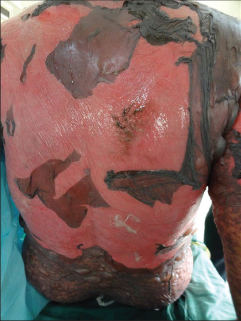
A 47-year-old woman, being treated for trigeminal neuralgia with tablet carbamazepine 100 mg thrice daily for 2 weeks, presented with complaints of skin rash and fever of 3 days duration. General examination revealed pulse rate of 124/min and temperature of 100°F. Dermatological examination revealed involvement of approximately 70% of body surface area with vesiculobullous lesions and erosions [Figure - 1]. Oral and vaginal mucosa showed erosions and crusting. Investigations on admission revealed polymorphonuclear leukocytosis, blood urea: 56 mg/dl, serum creatinine: 1.4 mg/dl, serum bicarbonate: 24 mmol/L and blood sugar: 186 mg/dl. The rest of the investigations, including chest X-ray, blood and urine culture, surface swab and culture was within normal limits. SCORTEN score on the day of admission and day 3 was assessed to be 4.
 |
| Figure 1: Widepread erosions |
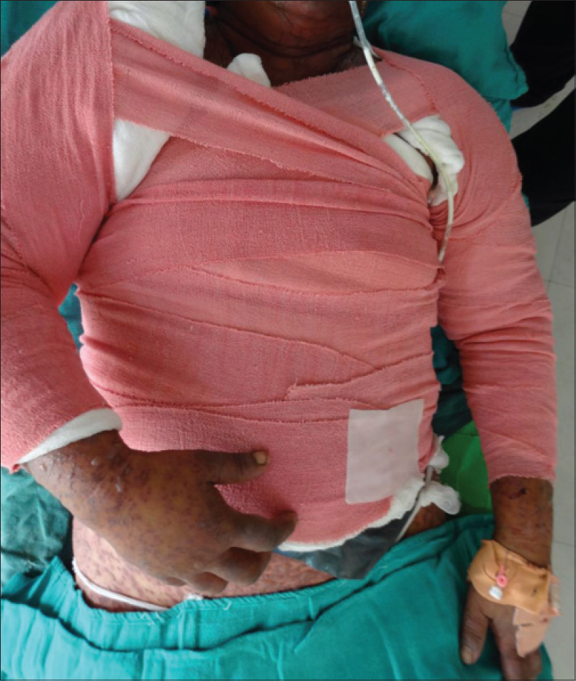
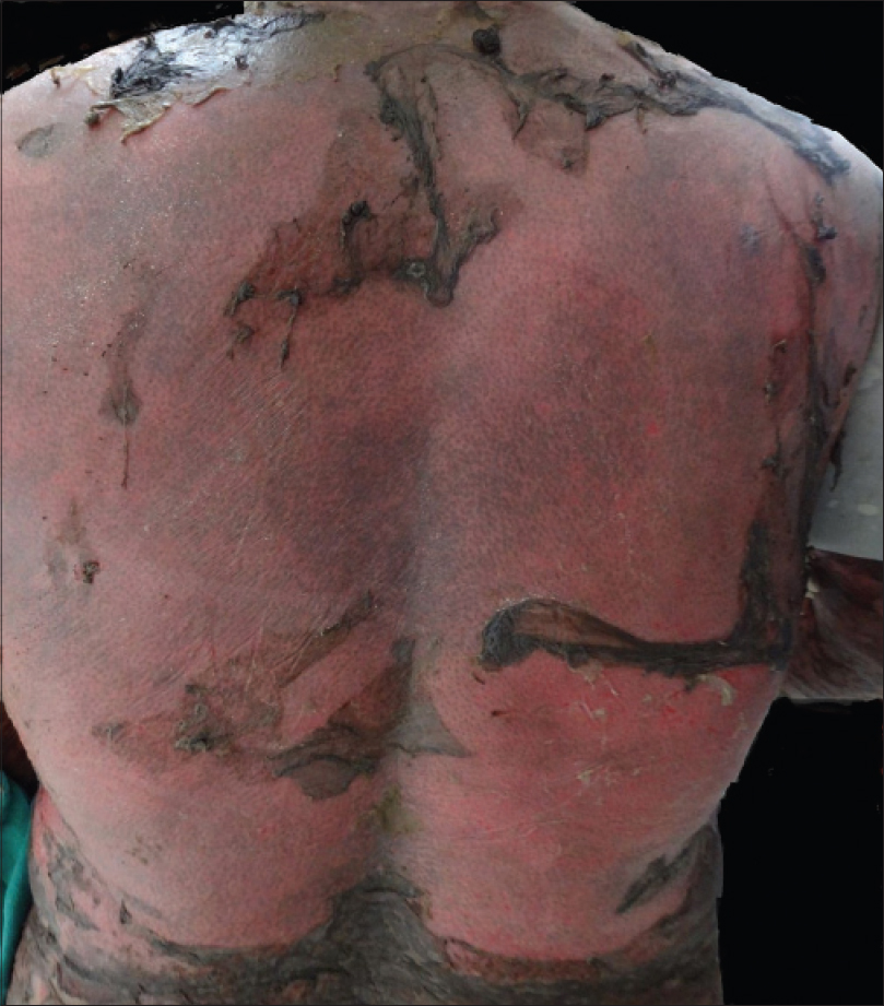
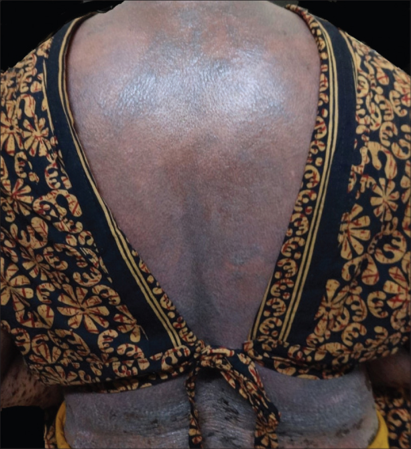
She was admitted to the burns ward and treated with intravenous immunoglobulin in a dose of 1 g/kg for 3 days along with supportive therapy. In view of the large body surface area involvement and high risk of sepsis, we decided to use nano-silver dressing for her skin care. As the skin swab and blood culture was negative, we did not use any empirical antibiotics. Nano-silver dressings were moistened with distilled water, applied on the skin and kept in place using cotton pad and crepe bandage [Figure - 2]. These dressings were changed twice at an interval of 2 days. Re-epithelialization was noted on day 5 and complete healing of skin was noted on day 9 of admission [Figure - 3] and [Figure - 4]. The patient became afebrile on day 4, ambulant on day 6 and was discharged on day 12 of admission.
 |
| Figure 2: Nano-silver dressing in situ with cotton pad and crepe bandage |
 |
| Figure 3: Beginning of re-epithelialisation on day 5 |
 |
| Figure 4: Complete re-epithelialisation on day 9 |
Prevention of wound infection in toxic epidermal necrolysis remains a priority in management.[3] Silver has antimicrobial and anti-inflammatory properties. It is biologically active in the soluble form and exerts an antimicrobial effect by blocking the cellular respiration of microorganisms, denaturing bacterial DNA and disrupting the cell membrane. Silver sulfadiazine was introduced in 1960 for the management of burn wounds and it reduced infections in burn wounds significantly. However, silver sulfadiazine cream has a short action, thus requiring frequent dressing change.[4] Use of nanotechnology in dressing leads to release clusters of extremely small and reactive silver particles. This results in a higher surface area of skin being in contact with the silver particles and better antimicrobial action.[5]
We used a proprietary three-layered dressing consisting of silver nanocrystals applied on rayon or polyester core (Acticoat ™). This dressing needs to be moistened with sterile water before being placed on the wound. Moistening not only releases up to 100 parts per million of ionized silver, but also provides moistened environment for wound healing. This dressing releases thirty times less silver cations as compared to the other forms of silver as silver sulfadiazine; however, these silver cations are more effective and have the property of sustained release.[6]
Complete re-epithelialization takes approximately 23 days with steroids and 16.7 days with cyclosporine; however, in our case, re-epithelialization occured in 9 days and patient was discharged on day 12.[7] Cyclosporine is advantageous as compared to intravenous immunoglobulin; however, we used intravenous immunoglobulin because of fear of infection in our case.[8] Various dressings have been used for the management of affected skin in toxic epidermal necrolysis including biological dressings such as collagen sheets, amniotic membrane and homograft skin. Collagen sheets also prevent bacterial migration and improve wound healing, however, unlike silver dressing, these do not have an antimicrobial action.[9] Traditional dressings such as paraffin gauze do not have antimicrobial action and adhere to the wound surface, thus change of dressing results in de-epithelialization and is painful.
Use of nano-silver dressing prevents infection in toxic epidermal necrolysis which may lead to faster wound healing. In addition, it is a non-adhesive dressing and does not lead to denudation of skin during the change of dressing. As the dressing needs to be changed after 48–72 hrs, the pain and discomfort of change of dressing is reduced.
Financial support and sponsorship
Nil.
Conflicts of interest
There are no conflicts of interest.
| 1. |
Bastuji-Garin S, Fouchard N, Bertocchi M, Roujeau JC, Revuz J, Wolkenstein P. SCORTEN: A severity-of-illness score for toxic epidermal necrolysis. J Invest Dermatol 2000;115:149-53.
[Google Scholar]
|
| 2. |
Suwarsa O, Yuwita W, Dharmadji HP, Sutedja E. Stevens-Johnson syndrome and toxic epidermal necrolysis in Dr. Hasan Sadikin General Hospital Bandung, Indonesia from 2009-2013. Asia Pac Allergy 2016;6:43-7.
[Google Scholar]
|
| 3. |
Daeschlein G. Antimicrobial and antiseptic strategies in wound management. Int Wound J 2013;10 Suppl 1:9-14.
[Google Scholar]
|
| 4. |
Hoffmann S. Silver sulfadiazine: An antibacterial agent for topical use in burns. A review of the literature. Scand J Plast Reconstr Surg 1984;18:119-26.
[Google Scholar]
|
| 5. |
Heggers J, Goodheart RE, Washington J, McCoy L, Carino E, et al. Therapeutic efficacy of three silver dressings in an infected animal model. Journal of Burn Care and Research. 2005 Jan 1;26(1):53-6.
[Google Scholar]
|
| 6. |
Fong J, Wood F. Nanocrystalline silver dressings in wound management: A review. Int J Nanomedicine 2006;1:441-9.
[Google Scholar]
|
| 7. |
Singh GK, Chatterjee M, Verma R. Cyclosporine in Stevens Johnson syndrome and toxic epidermal necrolysis and retrospective comparison with systemic corticosteroid. Indian J Dermatol Venereol Leprol 2013;79:686-92.
[Google Scholar]
|
| 8. |
Kirchhof MG, Miliszewski MA, Sikora S, Papp A, Dutz JP. Retrospective review of Stevens-Johnson syndrome/toxic epidermal necrolysis treatment comparing intravenous immunoglobulin with cyclosporine. J Am Acad Dermatol 2014;71:941-7.
[Google Scholar]
|
| 9. |
Bhattacharya S, Tripathi HN, Gupta V, Nigam B, Khanna A. Collagen sheet dressings for cutaneous lesions of toxic epidermal necrolysis. Indian J Plast Surg 2011;44:474-7.
[Google Scholar]
|
Fulltext Views
4,013
PDF downloads
2,087


