Generic selectors
Exact matches only
Search in title
Search in content
Post Type Selectors
Search in posts
Search in pages
Filter by Categories
15th National Conference of the IAOMFP, Chennai, 2006
Abstract
Abstracts from current literature
Acne in India: Guidelines for management - IAA Consensus Document
Addendum
Announcement
Art & Psychiatry
Article
Articles
Association Activities
Association Notes
Award Article
Book Review
Brief Report
Case Analysis
Case Letter
Case Letters
Case Notes
Case Report
Case Reports
Clinical and Laboratory Investigations
Clinical Article
Clinical Studies
Clinical Study
Commentary
Conference Oration
Conference Summary
Continuing Medical Education
Correspondence
Corrigendum
Cosmetic Dermatology
Cosmetology
Current Best Evidence
Current Issue
Current View
Derma Quest
Dermato Surgery
Dermatopathology
Dermatosurgery Specials
Dispensing Pearl
Do you know?
Drug Dialogues
e-IJDVL
Editor Speaks
Editorial
Editorial Remarks
Editorial Report
Editorial Report - 2007
Editorial report for 2004-2005
Errata
Erratum
Focus
Fourth All India Conference Programme
From Our Book Shelf
From the Desk of Chief Editor
General
Get Set for Net
Get set for the net
Guest Article
Guest Editorial
History
How I Manage?
IADVL Announcement
IADVL Announcements
IJDVL Awards
IJDVL AWARDS 2015
IJDVL Awards 2018
IJDVL Awards 2019
IJDVL Awards 2020
IJDVL International Awards 2018
Images in Clinical Practice
In Memorium
Inaugural Address
Index
Knowledge From World Contemporaries
Leprosy Section
Letter in Response to Previous Publication
Letter to Editor
Letter to the Editor
Letter to the Editor - Case Letter
Letter to the Editor - Letter in Response to Published Article
LETTER TO THE EDITOR - LETTERS IN RESPONSE TO PUBLISHED ARTICLES
Letter to the Editor - Observation Letter
Letter to the Editor - Study Letter
Letter to the Editor - Therapy Letter
Letter to the Editor: Articles in Response to Previously Published Articles
Letters in Response to Previous Publication
Letters to the Editor
Letters to the Editor - Letter in Response to Previously Published Articles
Letters to the Editor: Case Letters
Letters to the Editor: Letters in Response to Previously Published Articles
Medicolegal Window
Messages
Miscellaneous Letter
Musings
Net Case
Net case report
Net Image
Net Images
Net Letter
Net Quiz
Net Study
New Preparations
News
News & Views
Obituary
Observation Letter
Observation Letters
Oration
Original Article
ORIGINAL CONTRIBUTION
Original Contributions
Pattern of Skin Diseases
Pearls
Pediatric Dermatology
Pediatric Rounds
Perspective
Presedential Address
Presidential Address
Presidents Remarks
Quiz
Recommendations
Regret
Report
Report of chief editor
Report of Hon : Treasurer IADVL
Report of Hon. General Secretary IADVL
Research Methdology
Research Methodology
Resident page
Resident's Page
Resident’s Page
Residents' Corner
Residents' Corner
Residents' Page
Retraction
Review
Review Article
Review Articles
Reviewers 2022
Revision Corner
Self Assessment Programme
SEMINAR
Seminar: Chronic Arsenicosis in India
Seminar: HIV Infection
Short Communication
Short Communications
Short Report
Snippets
Special Article
Specialty Interface
Studies
Study Letter
Study Letters
Supplement-Photoprotection
Supplement-Psoriasis
Symposium - Contact Dermatitis
Symposium - Lasers
Symposium - Pediatric Dermatoses
Symposium - Psoriasis
Symposium - Vesicobullous Disorders
SYMPOSIUM - VITILIGO
Symposium Aesthetic Surgery
Symposium Dermatopathology
Symposium-Hair Disorders
Symposium-Nails Part I
Symposium-Nails-Part II
Systematic Review and Meta-Analysis
Systematic Reviews and Meta-analyses
Systematic Reviews and Meta-analysis
Tables
Technology
Therapeutic Guideline-IADVL
Therapeutic Guidelines
Therapeutic Guidelines - IADVL
Therapeutics
Therapy
Therapy Letter
Therapy Letters
View Point
Viewpoint
What’s new in Dermatology
View/Download PDF

Translate this page into:

Letter to the Editor
2013:79:2;242-243
doi: 10.4103/0378-6323.107649

Comparison of direct microscopic methods using potassium hydroxide, periodic acid Schiff, and calcofluor white with culture in the diagnosis of onychomycosis

Sachin Yadav1 , AK Saxena1 , Malini R Capoor2 , V Ramesh1
1 Department of Dermatology and STD, Safdarjang Hospital and VMMC, New Delhi, India
2 Department of Microbiology, Safdarjang Hospital and VMMC, New Delhi, India

Correspondence Address:
Sachin Yadav
Department of Dermatology and STD, Safdarjang Hospital and VMMC, New Delhi
India
How to cite this article:
Yadav S, Saxena A K, Capoor MR, Ramesh V. Comparison of direct microscopic methods using potassium hydroxide, periodic acid Schiff, and calcofluor white with culture in the diagnosis of onychomycosis. Indian J Dermatol Venereol Leprol 2013;79:242-243
Copyright: (C)2013 Indian Journal of Dermatology, Venereology, and Leprology

Sir,

Onychomycosis is the most common cause of abnormal nails (more than 50%). [1] Given the differential diagnosis of dystrophic nails, it is important to obtain a definitive diagnosis of fungal infection before initiation of therapy. Potassium hydroxide (KOH), which is routinely used for staining mycological specimens, often yields false-negative results. [2] It has been reported that periodic acid Schiff (PAS) and calcofluor white (CW) might be useful stains in diagnosing fungal elements with high positivity rate. [2],[3] The purpose of this study was to compare KOH preparation, KONCPA (KOH-treated nail clippings + periodic acid Schiff) technique, and fluorescent microscopy using CW, with culture in the diagnosis of onychomycosis.

An open prospective study was conducted on patients attending outpatient department of dermatology in our hospital, between November 2009 and October 2010. Patients with clinical features of onychomycosis were evaluated. Those who had taken systemic antifungal treatment within last 9 months or who did not demonstrate fungal elements in direct microscopy and/or culture, in repeat sample also, were excluded.

After disinfecting with 70% alcohol, nail scrapings and subungual debris were obtained from affected areas with the sterile no. 15 scalpel blade. In KOH mount, a portion of nail specimen was incubated in 20% KOH overnight at room temperature. The softened nail material was gently crushed with cover slip to form a thin film over a clean glass slide and was examined microscopically for fungal elements. In KONCPA method, specimens were treated with 20% KOH at 56°C for 30 min and washed with isotonic sodium chloride solution. Specimens were then centrifuged at 3000 rpm for 5 min and sediment was crushed to form a thin film over the slide. Then, the film was stained with PAS and visualized under microscope. In fluorescent microscopy, KOH treated nail material was stained with fluorescent brightener, CW, on a glass slide and fungal elements were examined under fluorescent microscope. The rest of nail specimens were inoculated on Sabouraud′s dextrose agar medium with 0.05% chloramphenicol, with and without 0.5% cycloheximide for 4 weeks. The growth was identified by its cultural characteristics on media and sporulation pattern on lactophenol cotton. For pathogenic non-dermatophyte mold, the diagnosis required presence of hyphae in direct microscopy and isolation of same mold in primary and repeat nail sample.

Sixty-two patients had at least one of the four diagnostic methods positive for the presence of fungal elements. The positive rates of KOH preparation, KONCPA, fluorescent microscopy, and culture were 66.1%, 61.3%, 79.0%, and 75.8%, respectively. Considering culture as gold standard, sensitivity, specificity, positive predictive value, and negative predictive value were calculated for each microscopic method. The results are shown in [Table - 1].

Table 1: Comparison of results of microscopic methods

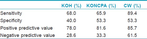
The common methods used for the diagnosis of onychomycosis are fungal culture on Sabouraud′s dextrose agar and KOH staining of nail sample. Culture was taken as gold standard test because of its high specificity and ability to identify species of invading pathogens. [2],[4],[5] The isolation rate of fungus on culture can be improved by taking enough nail sample from the most proximal affected area. KOH mount is a rapid, simple, and inexpensive method for diagnosis of fungal infections. The sensitivity of KOH preparations was 68% in this study. The different sensitivity rates reported earlier are 80%, [2] 55%, [4] 92%, [5] and 32%. [6] The false-negative results occur because of low visibility of scattered scanty fungal material obtained from the nail scraping and inadequate skill of observer in detection of fungal element. [4] Specificity of KOH preparations was found to be lower (40%). Artifacts including lipid vesicles, air bubbles, textile fibers, and mosaic fungus result in false-positivity. [7]

In KONCPA technique, PAS stain demonstrates the presence of certain polysaccharides, specifically glycogen and mucoproteins, as reddish structures which are present in the walls of the fungal hyphae. KONCPA showed sensitivity of 65.9% in our laboratory, which is lower than that reported in previous studies. [2],[4],[5] The lower detection was due to loss of major portion of KOH treated nail sample while washing with sodium chloride solution during the procedure. The false-positive results occur due to red staining of non-fungal structures like starch particles, serum remnants, and parakeratotic cells. [8]

Fluorescent microscopy showed high sensitivity of 89.4%, which was also noted in earlier reports. [2],[3],[5] This method is rapid and easy to perform. Fluorescent brighteners like CW specifically bind to cellulose and chitin, which are the major components in the cell wall of fungi. The dye then fluoresces as it is exposed to UV radiation on fluorescent microscopy. This helps in easy visualization of fungal elements. The constraint with this method is availability of fluorescent microscope and fluorescent dye. To conclude, the present study shows that CW microscopy gives significantly higher rates of detection of onychomycosis compared to the other microscopic methods using KOH and PAS stain.

References
1.
Welsh O, Vera-Cabrera L, Welsh E. Onychomycosis. Clin Dermatol 2010;28:151-9.
[Google Scholar]
2.
Weinberg JM, Koestenblatt EK, Tutrone WD, Tishler HR, Najarian L. Comparison of diagnostic methods in the evaluation of onychomycosis. J Am Acad Dermatol 2003;49:193-7.
[Google Scholar]
3.
Kwon KS, Yim CS, Jang HS, Oh CK, Chung TA. A Comparative Study for Diagnosing Onychomycosis Using KOH Smear, Fungal Culture, KONCPA and Fungi-Fluor(R) Stain. Korean J Med Mycol 1998;3:125-131.
[Google Scholar]
4.
Panasiti V, Borroni RG, Devirgiliis V, Rossi M, Fabbrizio L, Masciangelo R, et al. Comparison of diagnostic methods in the diagnosis of dermatomycoses and onychomycoses. Mycoses 2006;49:26-9.
[Google Scholar]
5.
Haghani I, Fathi M, Abedian S, Shokouhi T, Yazdani J. Comparison of diagnostic methods in the evaluation of onychomycosis. Cell J 2011;12:29-30.
[Google Scholar]
6.
Lawry MA, Haenke E, Strobeck K, Martin S, Zimmer B, Romano PS. Methods for diagnosing onychomycosis: A comparative study and review of literature. Arch Dermatol 2000;136:1112-6.
[Google Scholar]
7.
Baran R, Hay R, Haneke E, Tosti E, editors. Onychomycosis: The current approach to diagnosis and therapy. 2 nd ed. London: Informa Healthcare; 2006.
[Google Scholar]
8.
Pierard GE, Quatresooz P, Arresse JE. Spot light on histomycology. Dermatol Clin 2006;24:371-4.
[Google Scholar]

Fulltext Views
1,506

PDF downloads
230
Show Sections