Translate this page into:
Acrodermatitis enteropathica in three siblings
2 Department of Dermatology, Yuzuncu yil University, Faculty of Medicine, Van, Turkey
Correspondence Address:
Ayse Serap Karadag
Department of Dermatology, Istanbul Medeniyet University Faculty of Medicine, Goztepe Research and Training Hospital, Istanbul
Turkey
| How to cite this article: Karadag AS, Bilgili SG, Calka O. Acrodermatitis enteropathica in three siblings. Indian J Dermatol Venereol Leprol 2013;79:268 |
Sir,
Acrodermatitis enteropathica (AE) is a rare autosomal recessive disorder and occurs as a result of severe zinc deficiency in infants. It is caused by abnormal zinc absorption from the intestine. [1] Here, we reported three siblings with AE presenting with various degrees of skin lesions.
Case 1: A 12-year-old girl was admitted to our clinic for recurrent, erythtematous, scaly lesions on the face, arm, trunk, and perianal region. She had developed several episodes of lesions since the age of one. Hair loss and mouth soreness were accompanied with the lesions. The duration of lesions usually lasted for 10 days and healed without scar. The dermatological examination showed erythematous, xerotic, plaque-like, yellow-brown, dry, eczematous lesions in the perioral region [Figure - 1]a-b. The laboratory investigation showed low serum zinc level 66 μg/dl (N: 70-150 μg/dl)), and elevated alkaline 24 phosphates (776 U/l (N: 0-270 U/l)).
 |
| Figure 1: (a) Eczematous, erythematous lesions of perioral region of Case 1 (b) Appearance of facial lesions of Case 1-3 (c) Appearance post-treatment of Case 1-3 |
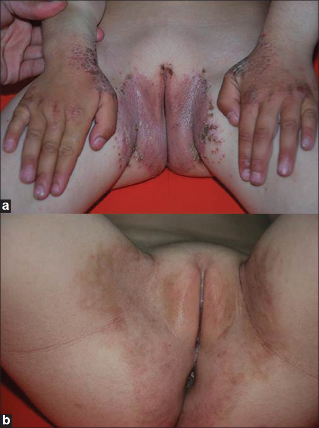
Case 2: A 6-year-old girl, sister of the Case 1 patient, presented with similar lesions localizing on her hands, arms, and body. She had had these lesions since she was aged 1 year. The lesions were accompanied with sore mouth and hair loss. Dermatological examination showed eroded and yellow-brown dry patches of plaques on the erythematous layer on the dorsum of the hand, perioral region, and inguinal region [Figure - 1]b and [Figure - 2]a.The laboratory tests showed low zinc (66.5 μg/dl) and elevated alkaline phosphatase level (428 U/l).
 |
| Figure 2: (a) Eczematous, erythematous lesions of perianal, intertriginous region of Case 2 (b) Appearance of perianal region post-treatment of Case 2 |
Case 3: A 3-year-old patient, brother of the Case 1 and 2 patients, presented with similar lesions from time to time. He had mild lesions in the perioral region. Histopathological examination of the lesions taken from the perianal region of case 2 showed parakeratosis associated with hypogranulosis, scattered dyskeratotic keratinocytes in the epidermis, and vacuolar degeneration of the basal layer. The findings were consistent with AE.
The patients′ parents were second-degree relatives (cousins). They had 5 siblings. The 3 patient were not initially examined by a dermatologist, but their primary care physician started them on a zinc treatment. Their lesions healed more quickly with zinc treatment so their family gave them zinc capsule whenever they developed new lesions. These patients (Cases 1-3) were treated with oral zinc sulfate capsule 50 mg/day and were instructed a life-long zinc treatment. The lesions disappeared within 2 weeks following the zinc treatment [Figure - 1]c and [Figure - 2]b.
AE can be acquired or congenital, and the prevalence of congenital AE is approximately 1 in 500,000 children. [2] In congenital AE, active zinc transport across the duodenal mucosa is defective, causing malabsorption and impaired uptake mechanism in the jejunal mucosa. The low zinc binding capacity of duodenal secretion suggested a defect in a transport facilitating ligand. In untreated AE cases, dietary zinc is not available for metabolism, and growth and whole body zinc stores are depleted; their tissue zinc concentration is low, zinc-dependent enzymes are inactive, and cell membranes are less stable. [3] In congenital forms, the AE gene, SLC39A43, is responsible for encoding the Zip4 zinc transporter. [1] The lesions in infants usually appear within days after birth in bottle-fed infants or after weaning from breast milk. However, infants on breastfeeding can still develop clinical zinc deficiency. [2] In our cases, there were no lesions during the breast-feeding period, and the lesions appeared at around the age of one and after staring additional food.
The reason of acquired zinc deficiency in children is prematurity, parenteral feeding, malnourishment, or cystic fibrosis. [2],[4] We assumed that our patients had congenital AE due to early onset of the lesions, no acquired additional diseases, and positive family history. However, we could not perform any further investigation to detect the exact mechanism of zinc malabsorption.
AE is characterized by a triad of acral and periorificial dermatitis, diarrhea, and alopecia; however, all three features are only present in around 20% of the cases. [1] The cutaneous lesion is usually the first sign of AE. AE presents as an erythematous, scaly, crusted, psoriasiform, eczematous, or vesiculobullous eruption around the body orifices on the extensor surfaces of major joints, acral sites, and the scalp. [1],[5] In mild hypozincemia, "scald like" erythema and lack of inflammation are the main features of the lesions. The lesions are accompanied with decreased hair and nail growth. [1],[4]
The diagnosis of AE can be made based on the clinical, histopathological findings, and laboratory abnormalities. A serum zinc level <50 mg/dl is indicative of AE, but not mandatory. AE can be seen in patients with normal zinc level. [4] Zinc-dependent enzymes such as alkaline phosphatase can be decreased. [2],[5] In our cases, zinc level was decreased and alkaline phosphates were elevated. Although laboratory tests are beneficial for diagnosis, zinc treatment responsiveness in suspected cases is the gold standard approach for the diagnosis. There is no clinical correlation between serum zinc level and severity of the disease. [1],[2] The AE patients require life-long zinc supplementation therapy to prevent further lesions. [1],[4]
| 1. |
Maverakis E, Fung MA, Lynch PJ, Draznin M, Michael DJ, Ruben B, et al. Acrodermatitis enteropathica and an overview of zinc metabolism. J Am Acad Dermatol 2007;56:116-24.
[Google Scholar]
|
| 2. |
Lee SY, Jung YJ, Oh TH, Choi EH. A case of acrodermatitis enteropathica localized on the hands and feet with a normal serum zinc level. Ann Dermatol 2011;23:S88-90.
[Google Scholar]
|
| 3. |
Van Wouwe JP. Clinical and laboratory diagnosis of acrodermatitis enteropathica. Eur J Pediatr 1989;149:2-8.
[Google Scholar]
|
| 4. |
Changela A, Javaiya H, Changela K, Davanos E, Rickenbach K. Acrodermatitis enteropathica during adequate enteral nutrition. JPEN J Parenter Enteral Nutr 2012;36:235-7.
[Google Scholar]
|
| 5. |
Jung AG, Mathony UA, Behre B, Küry S, Schmitt S, Zouboulis CC, et al. Acrodermatitis enteropathica: An uncommon differential diagnosis in childhood - first description of a new sequence variant. J Dtsch Dermatol Ges 2011;9:999-1002.
[Google Scholar]
|
Fulltext Views
2,263
PDF downloads
1,196


