Translate this page into:
A fleshy protuberant growth on the leg
2 Department of Pathology, National Institute of Pathology, Indian Council of Medical Research, New Delhi, India
Correspondence Address:
Sarvesh Sunil Thatte
Department of Dermatology, Venereology and Leprosy, Dr. P.N. Behl Skin Institute and School of Dermatology, New Delhi - 110 048
India
| How to cite this article: Bhushan P, Thatte SS, Singh A. A fleshy protuberant growth on the leg. Indian J Dermatol Venereol Leprol 2015;81:652-653 |
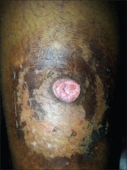
An 18-year-old boy presented with an asymptomatic, occasionally bleeding, flesh colored growth on his left leg for 10 years. A well-defined, 3 cm × 4 cm, flesh colored, sessile, soft, mobile, non-tender, nodule was noted on the left calf. The nodule protruded slightly from a shallow cup-shaped depression and had a slightly raised hyperpigmented rim. There was scaling and crusting of the surrounding skin because of repeated cleaning of the growth and surrounding area with undiluted 10% povidone-iodine solution [Figure - 1]. There were no other abnormalities on mucocutaneous examination.
 |
| Figure 1: Central mass projecting out of the cup-shaped moat |
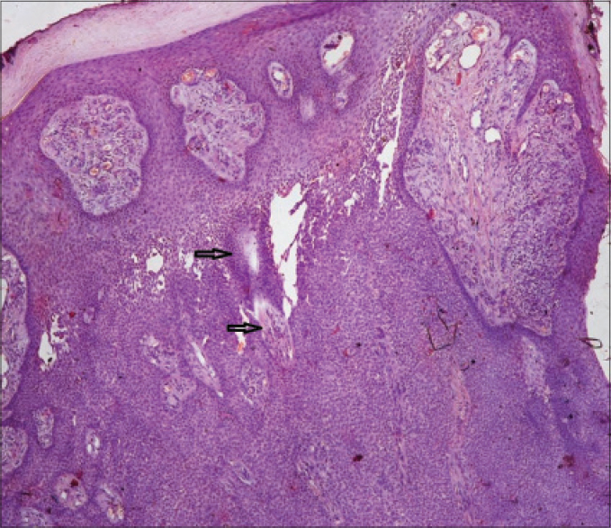
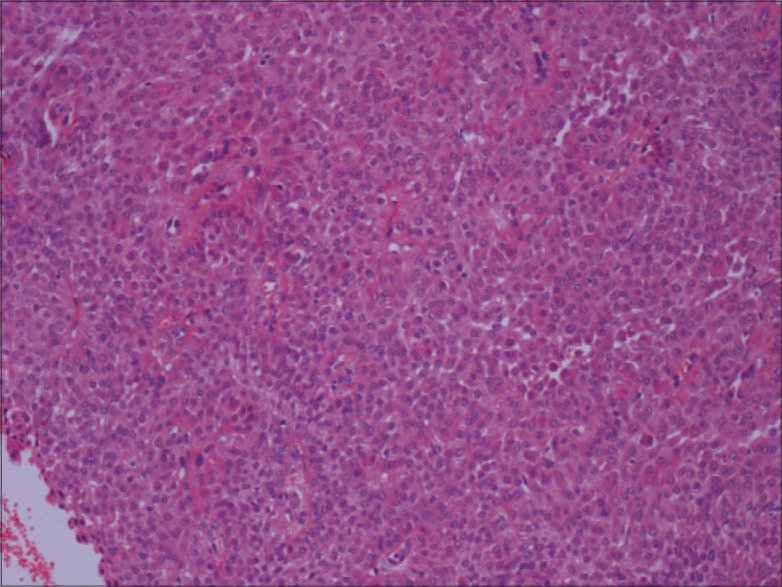
Histopathological examination revealed a well circumscribed tumor arising from the epidermis in the form of broad and anastomosing columns of basaloid cells along with necrosis en masse and a fibrovascular stroma [Figure - 2]. Medium power revealed small and uniform oval cells with a central nucleus, some ductal lumina and a fibrovascular core [Figure - 3].
 |
| Figure 2: A well circumscribed tumor arising from epidermis consisting of broad and anastomosing columns of basaloid cells along with necrosis en masse (arrow) (H and E, ×40) |
 |
| Figure 3: Small and uniform oval cells with central nucleus and some ductal lumina (H and E, ×100) |
What Is Your Diagnosis?
| 1. |
Srivastava D, Taylor RS. Appendage tumors and hamartomas of the skin. In: Goldsmith LA, Katz SI, Gilchrest BA, Paller AS, Leffell DJ, Wolf K, editors. Fitzpatrick's Dermatology in General Medicine. 8th ed. New York: McGraw-Hill; 2012. p. 1337-62.
[Google Scholar]
|
| 2. |
Agarwal S, Kumar B, Sharma N. Nodule on the chest. Eccrine poroma. Indian J Dermatol Venereol Leprol 2009;75:639.
[Google Scholar]
|
| 3. |
McCalmont TH. Adnexal neoplasms. In: Bolognia JL, Jorizzo JL, Schaffer JV, editors. Dermatology. 3rd ed. China: Elsevier Limited; 2012. p. 1829-49.
[Google Scholar]
|
| 4. |
Mantri MD, Dandale A, Dhurat RS, Ghate S. Pedunculated poroma on forearm: A rare clinical presentation. Indian Dermatol Online J 2014;5:469-71.
[Google Scholar]
|
| 5. |
Joshi RR, Nepal A, Ghimire A, Karki S. Eccrine poroma in neck of a child – A rare presentation. Nepal Med Coll J 2009;11:73-4.
[Google Scholar]
|
Fulltext Views
2,633
PDF downloads
1,667


