Translate this page into:
A tale of a reticulated erythematous patch on the breast
Corresponding author: Dr. Amal Chamli, Department of Dermatology, Habib Thameur Hospital, Tunis, Tunisia. amal.fmt@gmail.com
-
Received: ,
Accepted: ,
How to cite this article: Mrad M, Chamli A, Helal I, Khanchel F, Majdoub A, Fenniche S, et al. A tale of a reticulated erythematous patch on the breast. Indian J Dermatol Venereol Leprol 2023;89:867-9.
A 48-year-old woman, presented with a 3-month history of reticular erythematous poorly circumscribed patches over the inferior portion of her both breasts associated with recent painful ulceration on her right breast [Figure 1a and b]. Her body mass index was 36 and she had large pendulous breasts. She was a daily cigarette smoker with 30-pack-year history. Her past and family history was unremarkable. A biopsy from the reticular patch of the left breast and ulceration was done [Figures 2 and 3].
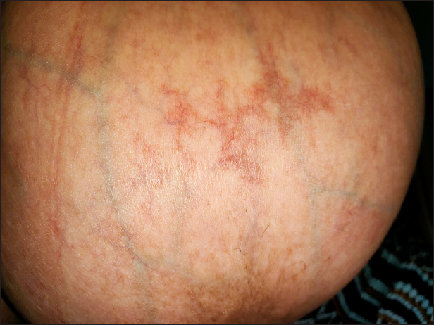
- Reticular erythematous plaques on the breast
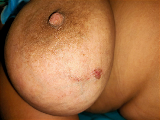
- Ulceration on the right side of the breast
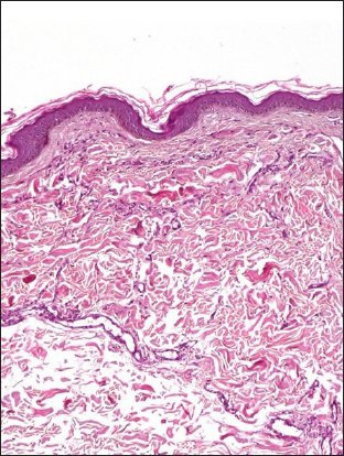
- The histopathological exam revealed a proliferation of capillary vessels with turgescent endothelial cells within the dermis, scattered extravasated erythrocytes with hemosiderin (HE × 100)
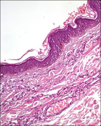
- Proliferation of capillary vessels within the dermis (HE × 200)
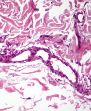
- High magnification shows proliferating endothelial cells and focal small vessel formation within the dermis (HE × 400)
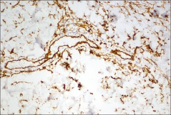
- Immunohistochemistry showed expression of CD34 highlighting the endothelial nature of proliferating cells within the dermis (staining CD34 × 200)
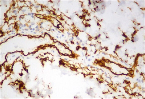
- Immunohistochemistry showed expression of CD34 highlighting the endothelial nature of proliferating cells within the dermis (staining CD34 × 400)
What is your diagnosis?
Diagnosis
Diffuse dermal angiomatosis of the breast
Discussion
The biopsy from the reticular plaque showed proliferation of vessels with turgescent endothelial cells within the dermis and scattered extravasated erythrocytes with hemosiderin [Figure 2]. There was no mitosis or vessel wall inflammation. Immunohistochemistry showed positive staining of endothelial cells for CD34. Staining for human herpesvirus 8 was negative. [Figure 3]. Laboratory investigations were within normal limits. Doppler ultrasound of subclavian arteries did not show any stenosis. Ultrasonic mammography revealed no mass or disrupted architecture. The patient was counselled to quit smoking and reduce weight. Reduction mammoplasty was suggested. Three months later, the ulcer was completely resolved & there was regression of breast patches. [Figure 4]. She had quit smoking, lost 11 kgs & started wearing an adapted bra which she wouldn’t have due to the unavailability of a fitting bra.
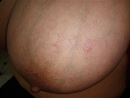
- Healing of the ulcer and net regression of the reticular patches over the breast
Diffuse dermal angiomatosis is a benign acquired vascular proliferation. It was recently recognised as a distinct rare variant of reactive angioendotheliomatosis with different pathological features.1,2 It is mostly located on the extremities but there is recently an increase in the reporting cases of the breast.2 It affects middle-aged women and clinically presents as reticulated, erythematous plaques that are usually multiple and bilateral. Superficial painful ulcerations may occur over the plaque, as in our case, and moreover, induration and necrosis of the plaque may also occur in some advanced cases with severe and chronic tissue hypoxia. Macromastia, obesity, subclavian artery stenosis, hypertension, hypercoagulable states, antiphospholipid syndrome and monoclonal gammopathy are associated with diffuse dermal angiomatosis of the breast.3,4 Chronic and focal hypoxia induces an up-regulation of vascular endothelial growth factors and stimulate angiogenesis.5 Subclinical torsion, compression, repeated microtrauma and increased pressure of large pendulous breasts contribute to chronic hypoxia of focal breast tissue.2,5 Furthermore, our patient and 27 % of described cases in the literature were heavy smokers.5 Indeed, smoking is known to cause endothelial dysfunction and a procoagulant state contributing to vascular damage and hypoxia.6
The distinctive histopathologic features of diffuse dermal angiomatosis of the breast are a diffuse proliferation of endothelial cells between collagen bundles, in contrast to the intraluminal proliferation of endothelial cells seen in classic reactive angioendotheliomatosis.4 The proliferating cells have a spindle-shaped appearance and vacuolated cytoplasm and form small vascular channels. Atypical endothelial cells and mitosis are absent. The pathological differential diagnoses include Kaposi sarcoma, post-radiation atypical vascular lesion, and low-grade angiosarcoma.7 Immunohistochemistry confirms the benign nature of endothelial proliferation. It shows positive vascular markers cluster of differentiation 31 and 34 (CD31, CD34), and Ets-related gene (ERG) with negative human herpesvirus 8 marker.3,8
Treatment depends on the underlying aetiology. Anti-aggregation may be beneficial for patients with hypercoagulability, especially in the presence of anti-cardiolipin and antinuclear antibodies. Aspirin and pentoxifylline were used and showed variable degrees of success.4,9 Furthermore, isotretinoin and corticosteroids have been described as an effective therapy in controlling the disease given their angiogenetic effects.5 Revascularisation of the subclavian artery has been described in cases associated with arterial obstruction. In addition, an excision of the painful and necrotic lesions may be proposed in severe cases.10 Reduction mammoplasty is proposed for patients with large pendulous breasts.3,5,10 The strict control of cardiovascular risk factors and smoking cessation is mandatory.3
Declaration of patient consent
Patient’s consent not required as patient’s identity is not disclosed or compromised.
Financial support and sponsorship
Nil.
Conflict of interest
There are no conflicts of interest.
References
- Cutaneous reactive angiomatoses: Patterns and classification of reactive vascular proliferation. J Am Acad Dermatol. 2003;49:887-96.
- [CrossRef] [PubMed] [Google Scholar]
- Diffuse dermal angiomatosis of the breast. Proc (Bayl Univ Med Cent). 2020;33:273-275.
- [CrossRef] [PubMed] [Google Scholar]
- Diffuse dermal angiomatosis of the breast: Clinical and histopathological features. Int J Dermatol. 2014;53:445-9.
- [CrossRef] [PubMed] [Google Scholar]
- Diffuse dermal angiomatosis of the breast: Clinicopathologic study of 5 patients. J Am Acad Dermatol. 2014;71:1212-7.
- [CrossRef] [PubMed] [Google Scholar]
- Diffuse dermal angiomatosis of the breast: A series of 22 cases from a single institution. Gland Surg. 2015;4:554-60.
- [CrossRef] [PubMed] [Google Scholar]
- Smoking and endothelial dysfunction. Curr Vasc Pharmacol. 2020;18:1-11.
- [CrossRef] [PubMed] [Google Scholar]
- Diffuse dermal angiomatosis of the breast: An emerging entity in the setting of cutaneous reactive angiomatoses. Clin Dermatol. 2021;39:271-7.
- [CrossRef] [PubMed] [Google Scholar]
- What is your diagnosis? Diffuse dermal angiomatosis secondary to anitcardiolipin antibodies. Am J Dermatopathol. 2002;24:502-3.
- [PubMed] [Google Scholar]
- Diffuse dermal angiomatosis of the breast with adjacent fat necrosis: A case report and review of the literature. Dermatol Online J. 2018;24 13030/qt1vq114n7
- [PubMed] [Google Scholar]


