Translate this page into:
Acrodermatitis enteropathica in a breast-fed infant
Correspondence Address:
Saurabh Agarwal
Department of Dermatology and Venereology, UFHT and Medical College Haldwani (Nainital) Uttaranchal-263 139
India
| How to cite this article: Agarwal S, Gopal K. Acrodermatitis enteropathica in a breast-fed infant. Indian J Dermatol Venereol Leprol 2007;73:209 |
 |
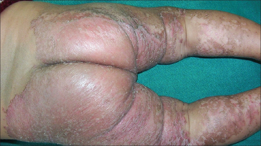
| Figure 1: Well-defi ned psoriasiform plaques over buttocks and legs |
 |
| Figure 1: Well-defi ned psoriasiform plaques over buttocks and legs |
Sir,
The term acrodermatitis enteropathica (AE) is now being used to include all patients with acral dermatitis due to zinc deficiency of hereditary or nonhereditary etiology. The hereditary form is an autosomal recessive disorder of zinc malabsorption seen exclusively in infants not receiving breast milk. Increased bioavailability of zinc from breast milk due to the presence of zinc-binding ligand confers protection of breast-fed infants against its deficiency. [1] However, acquired zinc deficiency may be seen rarely in premature or full-term breast-fed infants. [2] We describe a four-month old, breast-fed male infant with the typical acral rash of AE.
A 4-month-old male infant born normally at full-term to nonconsanguineous parents presented with a one-month history of rash over the face, hands, buttocks, legs and feet. No history of diarrhea or similar complaints was obtained. The rash had not responded to topical antifungal, topical corticosteroids, topical and systemic antibiotics prescribed elsewhere. The infant was exclusively breast-fed since birth and had not been weaned yet. General health and development of child was satisfactory except for poor weight gain in the last one month. Examination revealed an afebrile, active but irritable child weighing 4.2 kg with a head circumference of 40 cm. The general physical and systemic examination was unremarkable. On cutaneous examination, large erythematous, well-defined, psoriasiform, dry, scaly plaques of bilateral symmetrical distribution were present over the buttocks, thigh and legs [Figure - 1]. Flexures were relatively spared. Similar lesions were observed over the genital region, dorsum of fingers and toes and perioral region along with angular cheilitis. Scalp, hair, nails and mucosae were normal. A clinical diagnosis of AE was considered. Routine hematological and biochemical investigations were in normal limits. The child′s serum zinc level had decreased to 55 microgram/dl (normal: 70-120 microgram/dl). A microscopic examination of a potassium hydroxide (KOH) preparation of the skin scraping was negative. Treatment with oral zinc gluconate [5 mg/kg/day], was started and the parents were asked to start weaning. The skin lesions resolved in the next two weeks and the child became cheerful. There was no recurrence of symptoms / lesions on follow-up for two months.
AE is characterized clinically by a triad of dermatitis, diarrhea and alopecia although the complete triad is seen in only 20% of the patients. [3] The characteristic distribution of dermatitis over the face, hands, feet and anogenital region is recognized as a cutaneous marker of zinc deficiency. The cutaneous lesions are psoriasiform, erythematous, scaly and crusted plaques. As the disease progresses, these lesions may become vesicobullous, pustular and erosive. Other features, which occur with varying frequency, include stomatitis, apathy, irritability, growth retardation, failure to thrive and delayed wound healing. Delayed puberty and hypogonadism in developing males are some of the long-term effects of zinc deficiency. The ocular manifestations include photophobia, blepharitis, conjunctivitis and corneal dystrophy.
The actual metabolic error in AE has not yet been defined but it is clearly a problem with intestinal absorption and / or transport of zinc. In breast-fed infants with acquired zinc deficiency, maternal breast milk zinc levels are also reported to be low although maternal serum zinc levels may be normal. [2] This nonhereditary form of the disease develops due to low breast milk zinc levels secondary to defective mammary zinc secretion or an abnormal uptake of plasma zinc by the mammary glands despite normal maternal serum zinc levels. [4],[5] Treatment with 3-5 mg/kg/day zinc supplementation is recommended in children. There is a rapid improvement of diarrhea within 24 hours and of the skin lesions within 1-2 weeks. [1],[3] Prompt recognition of this disorder and initiation of zinc supplementation and weaning rapidly reverses the clinical features and helps to prevent the long-term consequences of zinc deficiency.
| 1. |
Sehgal VN, Jain S. Acrodermatitis enteropathica. Clin Dermatol 2000;18:745-8.
[Google Scholar]
|
| 2. |
Lee MG, Hong KT, Kim JJ. Transient symptomatic zinc deficiency in a full-term breast-fed infant. J Am Acad Dermatol 1990;23:375-9.
[Google Scholar]
|
| 3. |
Neldner KH. Acrodermatitis enteropathica and other zinc deficiency disorders. In : Fitzpatric TB, Freedberg IM, Eisen AZ, Wolff K, Austen KF, Goldsmith LA, Katz SI, editors. Dermatology in General Medicine 6 th ed. McGraw-Hill: New York; 2003. p. 1412-8.
[Google Scholar]
|
| 4. |
Zimmerman AW, Hambidge M, Lepow MI, Greenberg RD, Stover ML, Casey CE. Acrodermatitis in breast-fed premature infants: Evidence for a defect of mammary zinc secretion. Pediatrics 1982;69:176-83.
[Google Scholar]
|
| 5. |
Atkinson SA, Whelan D, Whyte RK, Lonnerdal B. Abnormal zinc content in human milk: Risk of development of nutritional zinc deficiency in infants. Am J Dis Child 1989;43:608-11.
[Google Scholar]
|
Fulltext Views
2,961
PDF downloads
3,381


