Translate this page into:
An asymptomatic inguinal swelling: Lymphatic filariasis
2 Department of Pathology, UCMS and GTB Hospital, New Delhi, India
Correspondence Address:
Amit Kumar Dhawan
Dr. Dhawan�s Skin, Cosmetology and Laser Clinic, House No. 436, Second Floor Indra Vihar, New Delhi - 110 009
India
| How to cite this article: Dhawan AK, Bisherwal K, Grover C, Sharma S. An asymptomatic inguinal swelling: Lymphatic filariasis. Indian J Dermatol Venereol Leprol 2016;82:446-448 |
Sir,
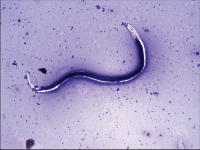
A 25-year-old labourer, from Uttar Pradesh presented with an asymptomatic gradually progressive swelling in the right inguinal region for 1 month. He had no history of urethral discharge, genital ulcer, any systemic symptoms or a similar previous episode. On examination, there was a single subcutaneous slightly tender, firm, mobile 3.5 × 3 cm swelling in the right inguinal region [Figure - 1]. There were no other findings on local and systemic examination. The patient underwent fine-needle aspiration cytological examination from the lesion with differential diagnoses of tubercular lymphadenitis, filariasis and inguinal bubo. On May-Grunwald-Giemsa staining of the smear, a few neutrophils and a filamentous organism suggestive of microfilaria was seen. The microfilaria had a peripheral sheath, was gently curved with a dark nuclear column in the body not extending to the tip of the tail [Figure - 2]. Hematological examination showed peripheral eosinophilia. Serology for the human deficiency virus (1 and 2) was non-reactive. Based upon morphological features and immunochromatography based antigen-assay, a diagnosis of filarial lymphadenitis caused by Wuchereria bancrofti was made. The patient was advised diethylcarbamazine, 6 mg/kg as a single dose resulting in resolution of the swelling within 4 weeks.
 |
| Figure 1: Asymptomatic lymph node swelling in right inguinal region |
 |
| Figure 2: Fine needle aspirate from lymph node showing a filamentous structure with peripheral sheath, dark nuclear column in its cavity and an empty space at the tip of its tail. (May-Grunwald-Giemsa stain, ×400) |
Lymphatic filariasis is endemic world-wide and nearly 1.4 billion people are estimated to be at risk globally. Asia, Africa, Pacific Islands, South America and Caribbean basin are the most affected geographic areas.[1] In India, the endemic states are Bihar with the highest endemicity (over 17%), Kerala (15.7%) and Uttar Pradesh (14.6%).[2] Wuchereria bancrofti, Brugia malayi, Onchocerca volvulus and Loa loa are the important filarial species and the vectors are Culex quinquefasciatus (W. bancrofti) and Mansonia annulifera (B. malayi).[1] Parasites are deposited in the human body by the bite of infected mosquitoes. The larvae then migrate to loco-regional lymph nodes and mature into adult forms. The mature fertilized female worm releases microfilariae into the peripheral circulation which are ingested by mosquitoes during a blood meal and develop into infective larvae in the mosquito over 10–14 days. These larvae migrate to the mosquito's proboscis and are inoculated in the human body perpetuating the cycle.[3],[4]
Adult filarial worms elicit a type 2 helper cell inflammatory response resulting in the elaboration of interleukins 4, 5, 9 and 10. This results in granuloma formation, endothelial and connective tissue proliferation, elevated IgE levels and peripheral blood and tissue eosinophilia. The irreversible anatomical and functional changes in lymphatic channels lead to dilatation and tortuousity of lymphatic vessels.[5]
Lymphatic filariasis can have varied clinical manifestations: acute lymphadenitis, acute lymphangitis, asymptomatic subcutaneous swelling, funiculo-epididymoorchitis, chronic lymphedema, hydrocele, lymph node swellings, nodules in breast, thyroid or salivary gland, tropical pulmonary eosinophilia and chyluria.[6],[7],[8] Lymphatic filariasis represents about 25–30% of all cases of filariasis. Conversely, filariasis accounts for 0.047% of all cases of lymph node swellings.[7] Recurrent episodes of lymphangitis predispose to chronic lymphedema and increased bacterial and fungal infections. Gradually, patients develop deformities affecting the quality of life.[6]
Non-invasive methods for diagnosis of lymphatic filariais include imaging (ultrasonography and magnetic resonance imaging) which demonstrates the continuous twirling motion of echogenic particles representing either adult filarial worms or microfilaria in scrotal lymphatics (filarial dance sign). Lymphoscintigraphy aids in demonstration of anatomical and functional abnormalities in lymphatic channels using radiolabeled dextran. Species can be identified by the use of immunochromatographic and deoxyribonucleic acid probes using polymerase chain reaction which are sensitive and specific methods for diagnosing different species of filariasis.[6] The demonstration of microfilariae is possible in peripheral venous blood smears sampled at midnight or after diethylcarbamazine provocation during the day. Fine-needle aspiration cytology offers an easy, inexpensive and quick method to demonstrate microfilariae and adult worms.[6],[7],[8] However, microfilaria in fine-needle aspiration cytology is an uncommon finding despite the high incidence of filariasis. A very low detection rate of microfilaria has been reported from superficial sites like breast lumps, lymph nodes, thyroid swellings, scrotal swellings and soft tissue swellings by some authors.[7],[8] The detection rate of microfilaria has been found to be only 0.078% of all samples from lymph node swellings.[7] Careful screening of fine-needle aspiration cytology smears is therefore required for demonstration of the parasite in aspirates. The different species of microfilariae can be differentiated based upon body morphology, nuclear characteristics, staining pattern, presence or absence of nuclei at the tip and nocturnal motility.[3],[6] Treatment modalities include diethylcarbamazine, 6 mg/kg single dose or daily for 12 days in divided doses, oral ivermectin 200 μg/kg, and albendazole 400 mg twice daily for 2 weeks.[1],[6] The management of chronic lymphedema is difficult. Limb elevation, local care, regular exercise, massage, intermittent pneumatic compression of the affected part and surgical procedures such as lymph node venous shunt, omentoplaxy, excisional surgery and skin grafting have been used with variable success.[6] In endemic countries, mass administration of a single annual dose of oral albendazole 400 mg and diethylcarbamazine 6 mg/kg of body weight is recommended for prevention of transmission of filariasis.[1],[6] Several subunit vaccines have been identified for prophylaxis, however, there are still no effective vaccines available.[9]
Financial support and sponsorship
Nil.
Conflicts of interest
There are no conflicts of interest.
| 1. |
WHO Lymphatic Filariasis; 2014. Available from: http://www.who.int/mediacentre/factsheets/fs102/en/. [Last accessed on 2014 Jul 11].
[Google Scholar]
|
| 2. |
Raju K, Jambulingam P, Sabesan S, Vanamail P. Lymphatic filariasis in India: Epidemiology and control measures. J Postgrad Med 2010;56:232-8.
[Google Scholar]
|
| 3. |
Bain O, Babayan S. Behaviour of filariae: Morphological and anatomical signatures of their life style within the arthropod and vertebrate hosts. Filaria J 2003;2:16.
[Google Scholar]
|
| 4. |
Simonsen PE, Mwakitalu ME. Urban lymphatic filariasis. Parasitol Res 2013;112:35-44.
[Google Scholar]
|
| 5. |
Babu S, Nutman TB. Immunopathogenesis of lymphatic filarial disease. Semin Immunopathol 2012;34:847-61.
[Google Scholar]
|
| 6. |
Palumbo E. Filariasis: Diagnosis, treatment and prevention. Acta Biomed 2008;79:106-9.
[Google Scholar]
|
| 7. |
Khare P, Kala P, Jha A, Chauhan N, Chand P. Incidental diagnosis of filariasis in superficial location by FNAC: A retrospective study of 10 years. J Clin Diagn Res 2014;8:FC05-8.
[Google Scholar]
|
| 8. |
Mitra SK, Mishra RK, Verma P. Cytological diagnosis of microfilariae in filariasis endemic areas of eastern Uttar Pradesh. J Cytol 2009;26:11-4.
[Google Scholar]
|
| 9. |
Dakshinamoorthy G, Samykutty AK, Munirathinam G, Reddy MV, Kalyanasundaram R. Multivalent fusion protein vaccine for lymphatic filariasis. Vaccine 2013;31:1616-22.
[Google Scholar]
|
Fulltext Views
6,022
PDF downloads
2,313


