Generic selectors
Exact matches only
Search in title
Search in content
Post Type Selectors
Search in posts
Search in pages
Filter by Categories
15th National Conference of the IAOMFP, Chennai, 2006
Abstract
Abstracts from current literature
Acne in India: Guidelines for management - IAA Consensus Document
Addendum
Announcement
Art & Psychiatry
Article
Articles
Association Activities
Association Notes
Award Article
Book Review
Brief Report
Case Analysis
Case Letter
Case Letters
Case Notes
Case Report
Case Reports
Clinical and Laboratory Investigations
Clinical Article
Clinical Studies
Clinical Study
Commentary
Conference Oration
Conference Summary
Continuing Medical Education
Correspondence
Corrigendum
Cosmetic Dermatology
Cosmetology
Current Best Evidence
Current Issue
Current View
Derma Quest
Dermato Surgery
Dermatopathology
Dermatosurgery Specials
Dispensing Pearl
Do you know?
Drug Dialogues
e-IJDVL
Editor Speaks
Editorial
Editorial Remarks
Editorial Report
Editorial Report - 2007
Editorial report for 2004-2005
Errata
Erratum
Focus
Fourth All India Conference Programme
From Our Book Shelf
From the Desk of Chief Editor
General
Get Set for Net
Get set for the net
Guest Article
Guest Editorial
History
How I Manage?
IADVL Announcement
IADVL Announcements
IJDVL Awards
IJDVL AWARDS 2015
IJDVL Awards 2018
IJDVL Awards 2019
IJDVL Awards 2020
IJDVL International Awards 2018
Images in Clinical Practice
Images in Dermatology
In Memorium
Inaugural Address
Index
Knowledge From World Contemporaries
Leprosy Section
Letter in Response to Previous Publication
Letter to Editor
Letter to the Editor
Letter to the Editor - Case Letter
Letter to the Editor - Letter in Response to Published Article
LETTER TO THE EDITOR - LETTERS IN RESPONSE TO PUBLISHED ARTICLES
Letter to the Editor - Observation Letter
Letter to the Editor - Study Letter
Letter to the Editor - Therapy Letter
Letter to the Editor: Articles in Response to Previously Published Articles
Letters in Response to Previous Publication
Letters to the Editor
Letters to the Editor - Letter in Response to Previously Published Articles
Letters to the Editor: Case Letters
Letters to the Editor: Letters in Response to Previously Published Articles
Media and news
Medicolegal Window
Messages
Miscellaneous Letter
Musings
Net Case
Net case report
Net Image
Net Images
Net Letter
Net Quiz
Net Study
New Preparations
News
News & Views
Obituary
Observation Letter
Observation Letters
Oration
Original Article
ORIGINAL CONTRIBUTION
Original Contributions
Pattern of Skin Diseases
Pearls
Pediatric Dermatology
Pediatric Rounds
Perspective
Presedential Address
Presidential Address
Presidents Remarks
Quiz
Recommendations
Regret
Report
Report of chief editor
Report of Hon : Treasurer IADVL
Report of Hon. General Secretary IADVL
Research Methdology
Research Methodology
Resident page
Resident's Page
Resident’s Page
Residents' Corner
Residents' Corner
Residents' Page
Retraction
Review
Review Article
Review Articles
Reviewers 2022
Revision Corner
Self Assessment Programme
SEMINAR
Seminar: Chronic Arsenicosis in India
Seminar: HIV Infection
Short Communication
Short Communications
Short Report
Snippets
Special Article
Specialty Interface
Studies
Study Letter
Study Letters
Supplement-Photoprotection
Supplement-Psoriasis
Symposium - Contact Dermatitis
Symposium - Lasers
Symposium - Pediatric Dermatoses
Symposium - Psoriasis
Symposium - Vesicobullous Disorders
SYMPOSIUM - VITILIGO
Symposium Aesthetic Surgery
Symposium Dermatopathology
Symposium-Hair Disorders
Symposium-Nails Part I
Symposium-Nails-Part II
Systematic Review and Meta-Analysis
Systematic Reviews and Meta-analyses
Systematic Reviews and Meta-analysis
Tables
Technology
Therapeutic Guideline-IADVL
Therapeutic Guidelines
Therapeutic Guidelines - IADVL
Therapeutics
Therapy
Therapy Letter
Therapy Letters
View Point
Viewpoint
What’s new in Dermatology
View/Download PDF

Translate this page into:

Letter to the Editor
2012:78:3;368-371
doi: 10.4103/0378-6323.95462

Correlating epidermal thickness and basement membrane length to angiogenesis in the centre and the periphery of vitiligo lesion

Voudouris Stylianos, Ioannidis Eleftherios, Kavantzas Nikolaos, Aroni Kyriaki
 Department of Dermatopathology, Medical School, National and Kapodistrian University of Athens, Athens, Greece

Correspondence Address:
Aroni Kyriaki
Mikras Asias 75 str, Goudi, Athens, 11527
Greece
How to cite this article:
Stylianos V, Eleftherios I, Nikolaos K, Kyriaki A. Correlating epidermal thickness and basement membrane length to angiogenesis in the centre and the periphery of vitiligo lesion. Indian J Dermatol Venereol Leprol 2012;78:368-371
Copyright: (C)2012 Indian Journal of Dermatology, Venereology, and Leprology

Sir,

In vitiligo it is well-documented that epidermis is thicker than in normally pigmented skin in frozen sections and that inflammatory changes are more frequent and intense in the epidermis and dermis of perilesional skin. Moreover angiogenesis, vascular endothelial growth factor (VEGF)-positive cells and mast cells are increased in the center compared to the periphery. [1],[2],[3]

Based on the above we investigated the thickness of keratin, the subjacent epithelium and the length of the basement membrane in the center (final stage) and the periphery (initial stage) of vitiligo, in order to find any difference between the two areas of the same lesion and a possible correlation between epidermal thickness and increased dermal angiogenesis.

After the approval of the Ethical Committee of the Medical School of the University of Athens biopsies were taken with informed consent from a group of 33 Greek patients (9 men, 24 women; age range, 20-72 years), with different phototypes [Fitzpatrick II (n=7), Fitzpatrick III (n=11), Fitzpatrick IV (n=15)], all suffering from generalized vitiligo vulgaris, stable for at least 2 years. The patients did not reveal any hyper- or hyposensitivity to pain, heat, cold, tickle, or touch, and did not show other abnormal sensations in their involved or clinically nondepigmented (uninvolved) skin areas. The duration of the disease ranged from 2 to 35 years. The patients had not received any treatment for vitiligo for the last 5 years atleast.

Biopsies (4 mm) were taken from the center and the periphery of the same lesion in each patient. Every effort was made to ensure that the diameter of tissue taken, the subsequent measurement of basement membrane length and the percentage of shrinkage of the tissue specimen after excision, due to skin elasticity would not be affected by the procedure. The thickness of keratin layer and subjacent epithelium (granular layer to basement membrane) was measured in H-E (Hematoxylin-Eosin)-stained sections (center and periphery). The length of the basal membrane was measured in PAS (Periodic Acid-Schiff)-stained sections.

All above measurements were performed semi-automatically. Specifically, for the measurement of the thickness were selected five different sites including the thicker and the thinner of each section. Then by the appropriate image analysis software (Image ProPlus, V.5, Media Cybernetics, Bethesda, MD, USA) the measurements were performed automatically. A grid of 4-mm long was used to perform accurate measurement of the full length of the basal membrane and cases with slides not properly embedded and cut and with different full length between center and periphery were excluded.

Endothelial cells were visualized using mouse monoclonal antibody against CD34 (Novocastra, Leika Microsystems, Newcastle upon Tune, UK).

The evaluation of histochemical and immunohistochemical stains was made by image analysis.

Statistical analysis was based on paired-t test.

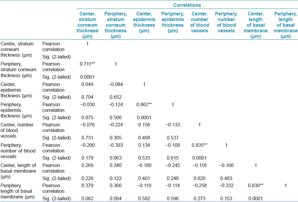
Results are demonstrated in [Table - 1], [Table - 2] and [Table - 3].
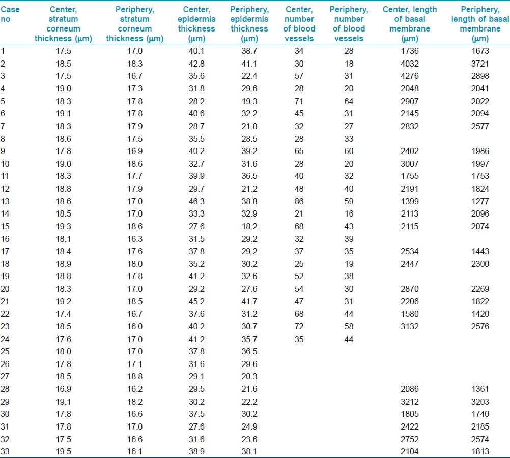
Table 1: Analytical results of the samples examined
Table 2: Statistical correlation between thickness of stratum corneum and epidermis, number of blood vessels and length of the epidermis in the center and the periphery of vitiligo lesion
Table 3: Percentage mean, median and standard deviation values of the thickness of stratum corneum and epidermis, the number of blood vessels and the length of the epidermis in the center and the periphery of vitiligo lesion

The increased thickness of the stratum corneum and the underlying epidermis and the loss of melanin were constant findings in all our cases, both in sun and non-sun-exposed sites, independently from the age of the lesion. In our study the thickness of stratum corneum and underlying epidermis in the center was higher, compared to the periphery of the lesion and basal membrane was longer in the center than in the periphery. Increased length of basement membrane is the result of increased number and/or length of epidermal rete ridges. All the above measurements allow us to conclude that the depigmented epidermis in vitiligo became hyperplasic with increased rete ridges.

It is well known that there are two routes of photoprotection, pigmentary and nonpigmentary route (increase of epidermal thickness). The last one is seen in vitiligo. [3] Epidermal hyperplasia in vitiligo occurs independently both in high and low sun exposed body areas. [1]

It has been reported that UV (Ultra Violet) exposure induces proliferation of the epidermal cells. Even a single exposure to UVB radiation can induce erythema, epidermal hyperplasia, and increased cutaneous vascularization. [4]

The increased number of dermal blood vessels in the center is also accompanied by hyperplasia of the overlying epidermis in 87% of our cases.

Epidermal hyperplasia and angiogenesis have an intimate relationship. Epithelial cells are dependent on the dermal blood supply during the phases of hyperproliferation. Evidence suggests that the stimulus for cutaneous neovascularization originates from epidermal, not dermal, cells. [4]

Angiogenesis accompanied by epidermal hyperplasia, is a histological finding observed also in psoriatic lesions. In psoriasis the morphological alterations of the blood vessels are prior to visible epidermal hyperplasia accompanied by lymphocytic infiltration. [5] In vitiligo there is not any known temporal correlation between increased angiogenesis and epidermal hyperplasia in the center of the lesion, where there is almost no lymphocytic infiltration. [2]

Factors like TNF-α (Tumor Necrosis Factor-alpha) and IL-6 (Interleukin-6) are increased in the center of vitiligo lesion compared to the periphery and can induce hyperplasia of the epidermis and increased number of blood vessels. [6]

In our study there wasn′t any linear correlation between these two parameters in the center of the lesion.

The loss of melanin leads to the above histopathological changes in the end stage of vitiligo, whereas its presence is important to maintain the normal cutaneous histological picture. This study, to the best of our knowledge, is the first to demonstrate the higher epidermal hyperplasia of the center compared to the periphery in vitiligo lesions, to correlate this epidermal hyperplasia to increased dermal angiogenesis and to attempt the measurement of basal membrane. Epidermal hyperplasia and accompanying increased angiogenesis may be part of a possible repairing/protective process. [7] We believe that the histological picture can help us understand disease′s pathogenesis better and lead us to choose the proper treatment.

References
1.
Gniadecka M, Wulf HC, Mortensen NN, Poulsen T. Photoprotection in vitiligo and normal skin. A quantitative assessment of the role of stratum corneum, viable epidermis and pigmentation. Acta Derm Venereol 1996;76:429-32.
[Google Scholar]
2.
Le Poole IC, van den Wijngaard R, Westerhof W, Das PK. Presence of T cells and macrophages in inflammatory vitiligo skin parallels melanocyte disappearance. Am J Pathol 1996;148:1219-28.
[Google Scholar]
3.
Aroni K, Voudouris S, Ioannidis E, Grapsa A, Kavantzas N, Patsouris E. Increased angiogenesis and mast cells in the centre compared to the periphery of vitiligo lesions. Arch Dermatol Res 2010;302:601-7.
[Google Scholar]
4.
Hennessy A, Oh C, Rees J, Diffey B. The photoadaptive response to ultraviolet exposure in human skin using ultraviolet spectrophotometry. Photodermatol Photoimmunol Photomed 2005;21:229-33.
[Google Scholar]
5.
Heidenreich R, Röcken M, Ghoreschi K. Angiogenesis drives psoriasis pathogenesis. Int J Exp Pathol 2009;90:232-48.
[Google Scholar]
6.
Kristensen M, Chu CQ, Eedy DJ, Feldmann M, Brennan FM, Breathnach SM. Localization of tumour necrosis factor-alpha (TNF-alpha) and its receptors in normal and psoriatic skin. Clin Exp Immunol 1993;94:354-62.
[Google Scholar]
7.
Bielenberg DR, Bucana CD, Sanchez R, Donawho CK, Kripke ML, Fidler IJ. Molecular regulation of UVB-induced cutaneous angiogenesis. J Invest Dermatol 1998;111:864-72.
[Google Scholar]

Fulltext Views
2,051

PDF downloads
1,158
Show Sections