Translate this page into:
Fibroepithelioma of Pinkus: A distinctive variant of trichoblastic carcinoma
Correspondence Address:
Rameshwar M Gutte
Department of Dermatology, OPD No. 112, 1st Floor, Dr. L. H. Hiranandani Hospital, Powai, Mumbai - 400 076, Maharashtra
India
| How to cite this article: Gutte RM. Fibroepithelioma of Pinkus: A distinctive variant of trichoblastic carcinoma. Indian J Dermatol Venereol Leprol 2013;79:725 |
Sir,
Fibroepithelioma of Pinkus (FEP) was initially described in 1953 by Pinkus, as premalignant fibroepithelial tumor of the skin. [1] Since then, FEP has been considered to be an unusual variant of basal cell carcinoma (BCC). However, some studies suggested that FEP is actually a variant of trichoblastoma and its classification still remains controversial. [2] Some consider it benign counterpart of BCC, while others think it a malignant neoplasm. [3]
Here, we report a case of FEP occurring in an adult male in association with nodular BCC, supporting its malignant nature.
A 40-year-old male, presented with an asymptomatic red colored nodule over the left side of his chest. Lesion had slowly grown to present size over a period of 5 years.
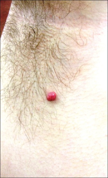
Examination revealed a solitary erythematous, non-tender sessile nodule of 0.6 mm × 0.8 mm size [Figure - 1]. No other skin abnormality was detected. Regional lymph nodes were not palpable.
 |
| Figure 1: Sessile pink colored nodule over the left side of chest |
A provisional diagnosis of pyogenic granuloma, Spitz nevus, appendageal tumor, and BCC was thought and excision with a wide margin was carried out under local anesthesia.
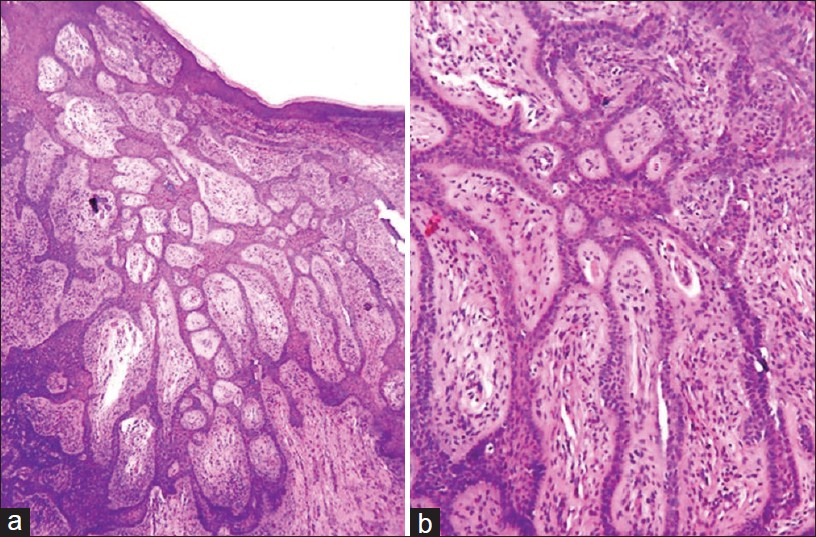
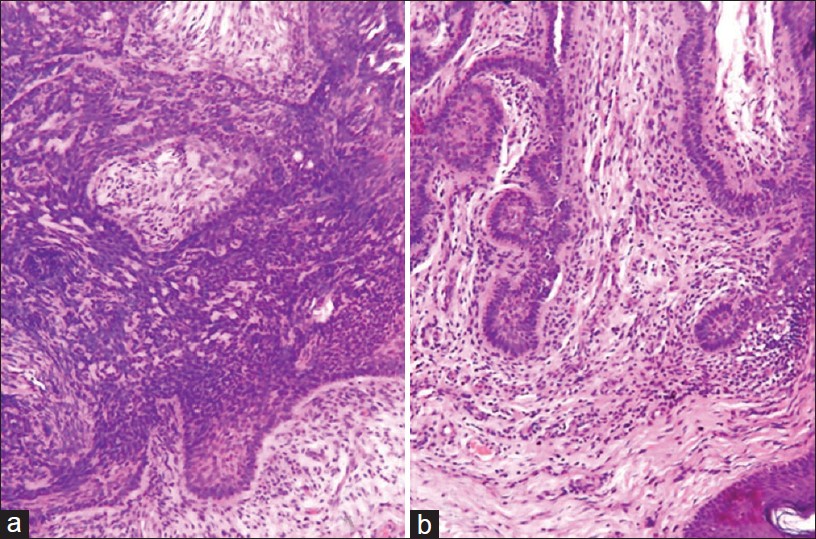
Histopathologic examination revealed an asymmetric neoplasm consisting of two different patterns produced by trichoblasts, namely, fenestrated, and nodular [Figure - 2]. Trichoblsats arranged in anastomosing epithelial struts separated by fibrotic stroma forming fenestrated pattern fulfilled criteria for fibroepithelial neoplasm [Figure - 3]. Because trichoblasts are also arranged in solid aggregations with crowding of cells, atypia, and peripheral palisading, criteria are met for nodular type of trichoblastic carcinoma. The struts being punctuated at places by protrusions forming follicular germ like structures unaffiliated with contiguous papilla, further qualifies it as trichoblastic neoplasm. The overall findings suggest FEP in continuity with a nodular BCC, a finding that supports the classification of FEP as a variant of BCC.
 |
| Figure 2: (a) Thin anastomosing strand of basaloid cells separated by fibrotic stroma along with solid aggregates on the left side (H and E, ×10). (b) High power view of anastomosing strands (H and E, ×40) |
 |
| Figure 3: (a) High power view of a nodular aggregate of trichoblasts with crowding, atypia and peripheral palisading (H and E, ×40). (b) Follicular germ-like structures (H and E, ×40) |
FEP usually presents with a single or multiple pedunculated or sessile nodules with a broad base on the trunk or extremities. In general, patients are male, 40-60 years of age, though the occurrence in children is recorded. [4] The tumor may appear pink, red, brown or skin color with occasional ulceration. Histologically, the tumor is composed of long, thin, and branching strands of basaloid cells anastomosing in fibrovascular stroma. [5]
Immunophenotypic evidence for the classification of FEP remains inconclusive. Similar to BCCs, FEP express androgen receptors. [6] In another study, authors found that tumor-specific type of epithelial hyperplasia in FEP that is positive for the stem cell marker PHLDA1 and present a unifying concept of FEP as a subtype of BCC. [7]
Local excision of FEP is recommended treatment and is almost universally curative. Neither aggressive behavior with local invasion nor metastasis has been reported. [5]
Our case shows the occurrence of nodular and fibroepithelial BCC in the same lesion and we suggest that what Pinkus called "premalignant fibroepithelial tumor" is not premalignant, but a malignant neoplasm made up of trichoblasts whose fenestrated design makes it distinctive among the various morphologic types of BCC.
| 1. |
Pinkus H. Premalignant fibroepithelial tumors of skin. AMA Arch Derm Syphilol 1953;67:598-615.
[Google Scholar]
|
| 2. |
Bowen AR, LeBoit PE. Fibroepithelioma of Pinkus is a fenestrated trichoblastoma. Am J Dermatopathol 2005;27:149-54.
[Google Scholar]
|
| 3. |
Ackerman AB, Gottlieb GJ. Fibroepithelial tumor of Pinkus is trichoblastic (Basal-cell) carcinoma. Am J Dermatopathol 2005;27:155-9.
[Google Scholar]
|
| 4. |
Pan Z, Huynh N, Sarma DP. Fibroepithelioma of Pinkus in a 9-year-old boy: A case report. Cases J 2008;1:21.
[Google Scholar]
|
| 5. |
Holcomb MJ, Motaparthi K, Grekin SJ, Rosen T. Fibroepithelioma of Pinkus induced by radiotherapy. Dermatol Online J 2012;18:5.
[Google Scholar]
|
| 6. |
Katona TM, Ravis SM, Perkins SM, Moores WB, Billings SD. Expression of androgen receptor by fibroepithelioma of Pinkus: Evidence supporting classification as a basal cell carcinoma variant? Am J Dermatopathol 2007;29:7-12.
[Google Scholar]
|
| 7. |
Sellheyer K, Nelson P, Kutzner H. Fibroepithelioma of Pinkus is a true basal cell carcinoma developing in association with a newly identified tumour-specific type of epidermal hyperplasia. Br J Dermatol 2012;166:88-97.
[Google Scholar]
|
Fulltext Views
4,793
PDF downloads
2,216


