Translate this page into:
Intratherapeutic dermoscopy assists nevus removal by laser therapy
Correspondence Address:
Chun-Yu Cheng
Chang Gung Memorial Hospital, 199, Tun-Hwa North Road, Taipei 105
Taiwan
| How to cite this article: Cheng CY. Intratherapeutic dermoscopy assists nevus removal by laser therapy. Indian J Dermatol Venereol Leprol 2020;86:604-605 |
Problem
Acquired melanocytic nevi are common benign pigmented cutaneous neoplasms. Traditional treatment of acquired melanocytic nevi includes dermabrasion, electrodesiccation and surgical excision. During recent years, laser therapy has become increasingly popular for the removal of acquired melanocytic nevi. Erbium-doped yttrium aluminum garnet laser emits a wavelength of 2,940 nm which is absorbed by tissue water. It can ablate and remove the superficial cutaneous lesions.[1] Although erbium-doped yttrium aluminum garnet laser is considered an effective and safe method for treatment of acquired melanocytic nevi, the therapeutic depth is often difficult to control.[2] Moreover, if depth of the treated area is not sufficient, the nevus would recur soon after treatment. On the other hand, scar formation would develop if the treated depth is more. Therefore, we have tried to find a solution to improve the outcome of laser procedure.
Solution
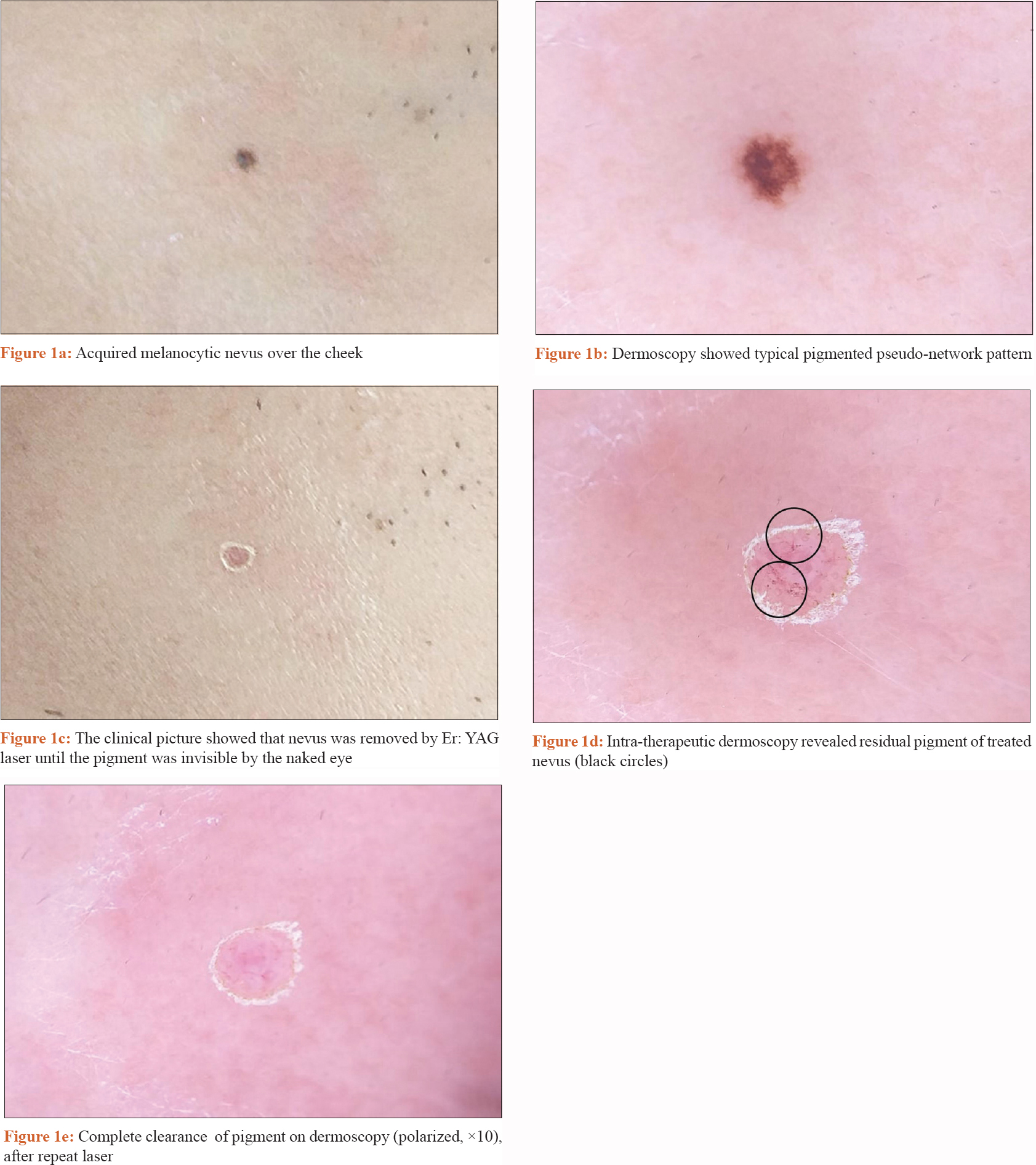
Dermoscopy is a non-invasive technique which has been used for assisting the diagnosis of pigmented or nonpigmented skin tumors for decades. We performed intra-therapeutic dermoscopy examination to enhance the clearance of melanocytic nevi by laser therapy. At first, the acquired melanocytic nevus [Figure - 1]a was examined by polarized dermoscopy (DermLite® DL4, 3 Gen, San Juan Capistrano, CA, USA) to rule out possible malignancy [Figure - 1]b. After adequate topical anesthesia, we used erbium-doped yttrium aluminum garnet laser (Profile, Sciton, Inc, Palo Alto, CA, USA) with a 2 mm spot size and fluency of 2.5 to 5.0 J/cm[2] to remove the nevus till the pigment was invisible by naked eye [Figure - 1]c. Later, we performed intra-therapeutic dermoscopy examination with non-contact polarized mode, to detect and map the residual pigment in the treated area [Figure - 1]d. Before dermoscopic examination, the contact plate was removed and the internal lens was cleaned using 70% alcohol. We also maintained 1 inch distance from the treated area to prevent contamination during the dermoscopic examination. Thereafter, we used erbium-doped yttrium aluminum garnet laser to treat the pigmented area, based on pigment mapping in dermoscopy for one to two passes. We repeated the process until there was no more pigment, as shown in dermoscopy examination [Figure - 1]e. After treatment, the wound was covered with hydrocolloid dressings for post-therapeutic care. The patient was followed up for at least 6 months without recurrence or scar formation.
 |
In conclusion, intra-therapeutic dermoscopy examination is a simple and non-invasive technique which can help us detect and map the location of residual nevus during laser therapy. In this way, we can remove the melanocytic nevus more accurately and avoid the damage of nearby tissue thereby reducing the risk of scar formation.
Declaration of patient consent
The authors certify that they have obtained all appropriate patient consent forms. In the form, the patients have given their consent for their images and other clinical information to be reported in the journal. The patient understands that name and initials will not be published and due efforts will be made to conceal identity but anonymity cannot be guaranteed.
Financial support and sponsorship
Nil.
Conflicts of interest
There are no conflicts of interest.
| 1. |
Arora H, Falto-Aizpurua L, Chacon A, Griffith RD, Nouri K. Lasers for nevi: A review. Lasers Med Sci 2015;30:1991-2001.
[Google Scholar]
|
| 2. |
Baba M, Bal N. Efficacy and safety of the short-pulse erbium: YAG laser in the treatment of acquired melanocytic nevi. Dermatol Surg 2006;32:256-60.
[Google Scholar]
|
Fulltext Views
4,021
PDF downloads
3,346


