Translate this page into:
Keratoacanthoma arising within a linear epidermal nevus
Correspondence Address:
Noureddine Litaiem
Department of Dermatology, Charles Nicolle Hospital, University of Tunis El Manar, Boulevard, 9 Avril 1938, 1006, Tunis
Tunisia
| How to cite this article: Litaiem N, Toumi A, Zeglaoui F. Keratoacanthoma arising within a linear epidermal nevus. Indian J Dermatol Venereol Leprol 2020;86:531-532 |
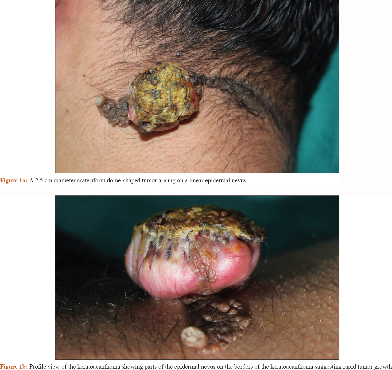
A 22-year-old male with an existing linear epidermal nevus on the nape of his neck, was referred for a dermatological evaluation of a rapidly growing nodule (of 3 weeks duration) that had developed within the epidermal nevus. On physical examination, there was a 2.5 cm diameter dome-shaped tumor [Figure - 1]a. Parts of the epidermal nevus were seen at the borders of the tumor [Figure - 1]b. A shave excision was performed to remove the tumor. Histopathological examination revealed symmetrical crateriform architecture with a central keratin plug and a peripheral squamous epithelial proliferation. On the basis of these clinico-pathological findings, the diagnosis of keratoacanthoma arising from a linear epidermal nevus was made. No relapses were noted, even at 2 months after removal.
 |
Mesh keywords
Keratoacanthoma, epidermal nevus, squamous cell carcinoma.
Declaration of patient consent
The authors certify that they have obtained all appropriate patient consent forms. In the form, the patient has given his consent for his images and other clinical information to be reported in the journal. The patient understands that name and initials will not be published and due efforts will be made to conceal identity, but anonymity cannot be guaranteed.
Financial support and sponsorship
Nil.
Conflicts of interest
There are no conflicts of interest.
Fulltext Views
3,113
PDF downloads
2,973


