Translate this page into:
Lupus vulgaris masquerading as tumorous growth
Corresponding author: Dr. Chandra Sekhar Sirka, Department of Dermatology and Venereology, AIIMS, Bhubaneswar, Sijua, Patrapada - 751 019, Odisha, India. csirka2006@gmail.com
-
Received: ,
Accepted: ,
How to cite this article: Sirka CS, Rout AN, Kumar P, Purkait S. Lupus vulgaris masquerading as tumorous growth. Indian J Dermatol Venereol Leprol 2021;87:562-5.
Sir,
Lupus vulgaris is the most common form of cutaneous tuberculosis in adults in India. Various clinical forms described include plaque-like, ulcerative, hypertrophic, vegetative, papular and nodular forms.1 Tumor-like and bulbous lesions have been described rarely in the literature.2-8 We report a case of lupus vulgaris which had resulted in tumorous transformation and distortion of the toes, causing confusion and delay in diagnosis and treatment leading to chronicity of the lesion and deformity.
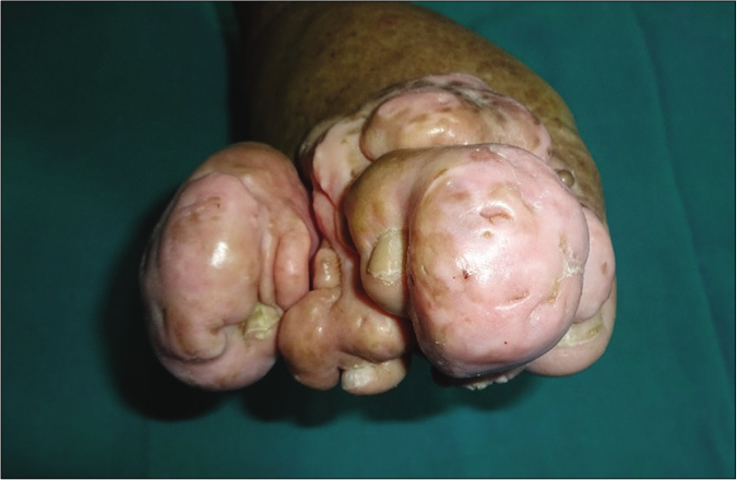
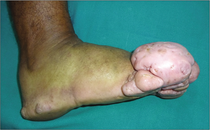
A 47-year-old male cook was referred to our outpatient department with tumor-like swellings on the distal right foot for 15 years. There was no history of trauma to the limb or any history of tuberculosis among close contacts. He had taken various medications with no improvement. The lesion started as redness and swelling of his second toe initially, following which in a few months, the toe got deformed and areas of depigmentation and verrucosity were visible [Figure 1a]. The lesions on the other parts started as nodules which enlarged and coalesced to form larger plaques [Figures 1b and c]. The bulbous swelling of the toes with a narrow base produced a pedunculated appearance. Few plaques spontaneously healed over a few months leaving atrophic and puckered scars [Figure 1a]. There were no palpable lymph nodes or any systemic symptoms. Based on these clinical features and evolution, possibilities of lupus vulgaris, chromoblastomycosis and cutaneous malignancy were considered.
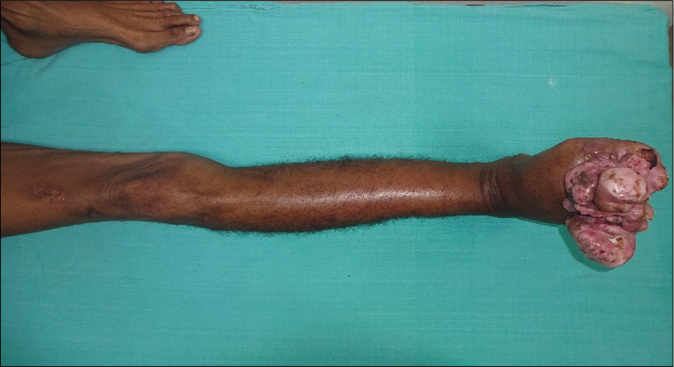
- Gross swelling with the fleshy tumor-like transformation of the toes
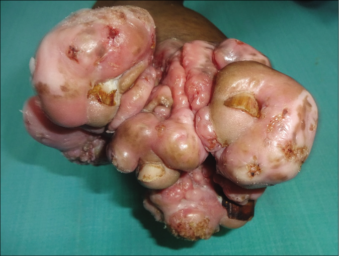
- Surface of the toes showing areas of verrucosity; nails visible on each of the toe
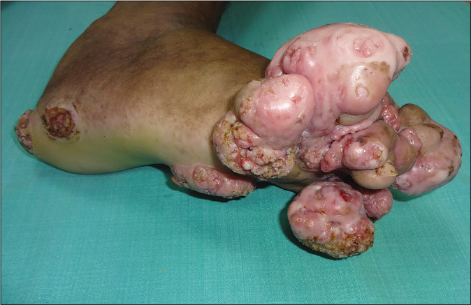
- Lateral view showing verrucosity on the surface of the nodular lesions and erythematous plaques with verrucosity
Hematological tests, renal and liver function tests were within normal limits. Screening for HIV was negative. The Mantoux test was positive (18 mm). X-ray of the chest was normal. X-ray of the feet showed distorted phalanx bones [Figure 1d]. The Erythrocyte sedimentation rate was 60 mm in 1st h. Biopsy from the lesion showed features suggestive of lupus vulgaris [Figures 2a-c]. Based on the clinical and laboratory findings, he was started on antitubercular therapy. After 6 months, there was an improvement in the verrucous part but leg swelling with smooth scars persisted [Figures 3a-c]. He was referred to the department of plastic surgery for reconstructive surgery.
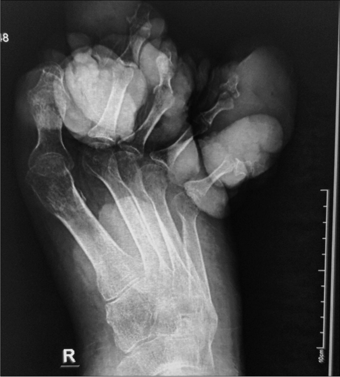
- X-ray of the right foot showing grossly distorted toes bones
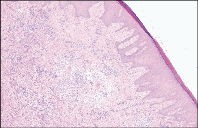
- Scanner view showing hyperkeratotic and acanthotic epidermis and the presence of multiple granulomas in deep dermis (H&E, ×40)
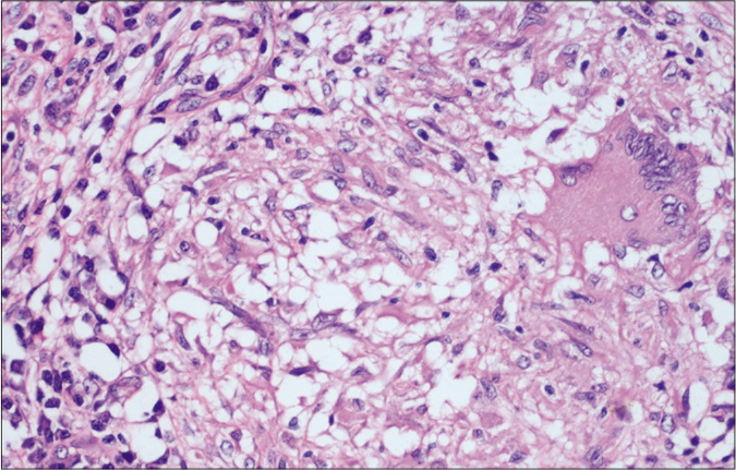
- High power view showing epithelioid cell granuloma with Langhans type giant cells and lymphohistiocytic infiltration in the surrounding area (H&E, ×400)
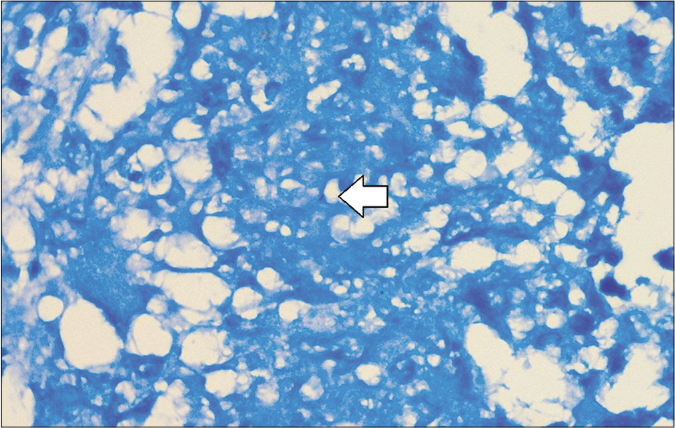
- Modified Ziehl–Neelsen stain showing acid-fast bacilli (Modified ZN, ×1000)
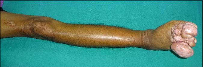
- Healing in the form of complete improvement of the surface verrucosity
- Healing in the form of complete improvement of the surface verrucosity
- Healing in the form of complete improvement of the surface verrucosity
The morphological presentation of lupus vulgaris is variable, thus, causing a diagnostic dilemma many times. The rare presentations include nodular, vegetating and papular forms.1
However, lupus vulgaris presenting as tumor-like and bulbous lesions is rare.2-8 [Table 1] In all the previously reported cases, tumor-like lesions were studded over the plaques of lupus vulgaris lesions, whereas in the present case, swelling and distortion of the toes had resulted in a nodular and bulbous look; which is evident from the presence of nail in each tumor-like growth and X-ray showing distorted phalanges and the toe bones.
| Serial number | Authors | Morphology |
|---|---|---|
| 1 | Garg et al.2 | 28-year female shiny erythematous plaque 3×2 cm with central atrophy and scarring on the face Multiple shiny nontender soft papules arranged in annular configuration discrete papules and nodules with adherent fine scaling bilaterally on the Alar prominence of the nose, lower lip and postauricular area Diascopy apple jelly nodule |
| 2 | Pilani et al.3 | 28-year-old female, laborer Progressive annular plaque over the right side of cheek extending up to right lower lid and ala of nose Two satellite plaques near the right side of giant lesion Diascopy apple jelly nodule |
| 3 | Lu et al.4 | 47 years female Massively enlarged earlobe with bluish-red or violaceous indurated plaques and nodules, with edema and ulceration |
| 4 | Gunawan et al.5 | 15 years female Erythematous plaque on the cheek and erythematous nodule on the index finger of the left hand Diascopy test(“apple jelly” sign): negative Multifocal skeletal TB(vertebrae and knee joint) |
| 5 | Hruza et al.6 | 69-year-old male Scattered, grouped, asymptomatic follicular papules, pustules, and nodules tending toward coalescence into large geographic aggregates |
| 6 | Kempter et al.7 | 61-year-old patient 25-year history of erythematous scaling lesions, wrongly diagnosed and treated as psoriasis vulgaris Nodular growth within the erythematous plaque |
| 7 | Bräuninger et al.8 | 74 years female Nodular infiltrations and confluating tumors |
TB: tuberculous
Diagnosis is mostly confirmed from raised erythrocyte sedimentation rate, lymphocytosis, positive Mantoux test and histology suggestive of tuberculoid granuloma. In the present case, the biopsies from the plaques and nodular looking lesion were suggestive of tuberculosis and he had raised erythrocyte sedimentation rate and positive Mantoux test.
Morphological differentials include squamous cell carcinoma, chromoblastomycosis and mycosis fungoides; however, the presence of tuberculoid granuloma on the biopsy can differentiate lupus vulgaris from other conditions.
A therapeutic trial of triple antituberculosis therapy: isoniazid, rifampicin and pyrazinamide may be considered in cases where the diagnosis is difficult. A clinical response would be expected within 4–6 weeks.9 In our case also the lesions showed improvement in the verrucous parts in 6 months.
We report a case of lupus vulgaris which had resulted in tumorous transformation and distortion of the toes, which had caused confusion and delay in diagnosis and led to the chronicity of the lesion and deformity, even in today’s era.
From the present case, we would like to suggest that lupus vulgaris on feet may masquerade as a tumor due to swelling and deformity of the toes.
Declaration of patient consent
The authors certify that they have obtained all appropriate patient consent.
Financial support and sponsorship
Nil.
Conflicts of interest
There are no conflicts of interest.
References
- A clinico-histopathological study of lupus vulgaris: A 3 year experience at a tertiary care centre. Indian Dermatol Online J. 2014;5:461-5.
- [CrossRef] [PubMed] [Google Scholar]
- Bilateral “turkey ear” as a cutaneous manifestation of lupus vulgaris. Indian J Dermatol Venereol Leprol. 2018;84:687-9.
- [CrossRef] [PubMed] [Google Scholar]
- A rare case of multiple lupus vulgaris in a multifocal tuberculosis pediatric patient. Int J Mycobacteriol. 2019;8:205-7.
- [Google Scholar]
- Disseminated lupus vulgaris presenting as granulomatous folliculitis. Int J Dermatol. 1989;28:388-92.
- [CrossRef] [PubMed] [Google Scholar]
- Therapy-resistant “psoriasis vulgaris”. Hautarzt. 2009;60:332-5.
- [CrossRef] [PubMed] [Google Scholar]
- How soon does cutaneous tuberculosis respond to treatment? Implications for a therapeutic test of diagnosis. Int J Dermatol. 2005;44:121-4.
- [CrossRef] [PubMed] [Google Scholar]


