Translate this page into:
Nicolau's syndrome following diclofenac administration: A report of two cases
Correspondence Address:
S Chidambara Murthy
Department of Dermatology and Venereology, Vijayanagara Institute of Medical Sciences, Bellary - 583 104, Karnataka
India
| How to cite this article: Murthy S C, Siddalingappa K, Suresh T. Nicolau's syndrome following diclofenac administration: A report of two cases. Indian J Dermatol Venereol Leprol 2007;73:429-431 |
 |
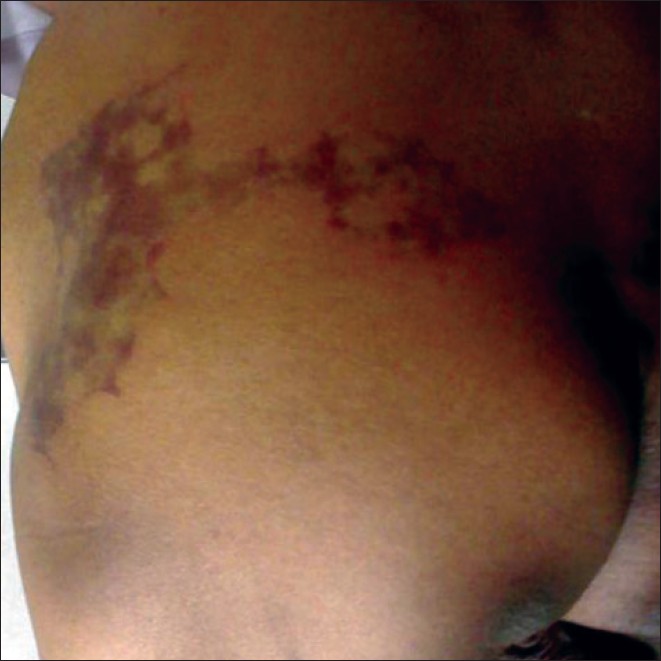
| Figure 2: Livedoid discoloration over the left gluteal region (Case 2) |
 |
| Figure 2: Livedoid discoloration over the left gluteal region (Case 2) |
 |
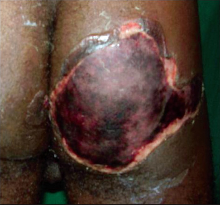
| Figure 1: Large deep ulcer over the right gluteal region with necrotic eschar in Case 1 |
 |
| Figure 1: Large deep ulcer over the right gluteal region with necrotic eschar in Case 1 |
Sir,
Nicolau′s syndrome (NS) is a rare injection site reaction, following intramuscular administration of drugs, with varying degrees of tissue damage. It is also synonymously described as embolia cutis medicamentosa [1] and livedoid dermatitis. [2] NS is characterized by development of an acute, severe pain and a localized erythematous rash following intramuscular injection. Subsequently cutaneous, subcutaneous and even muscular necrosis with a pale marble-like livedoid pattern results. [2] We report here two cases of NS following diclofenac administration.
Our first case was a 29 year-old man admitted with a history of snakebite, referred from the medicine department for evaluation of a painful ulcer over the right gluteal region. He had received two intravenous infusions of antivenin and an intramuscular (intragluteal) injection of diclofenac. After receiving diclofenac, he immediately noticed severe pain followed by blistering and ulceration.
Cutaneous examination showed a large tender, nonindurated ulcer with necrotic eschar covering almost the entire right gluteal region with minimal extension to the left [Figure - 1]. There was no regional lymphadenopathy. Other cutaneous and systemic examinations were normal. Complete hemogram including bleeding time, clotting time and urine examinations were normal. Chest X-ray, blood urea, serum creatinine, liver function tests, creatine kinase, were normal. Venereal disease research laboratory (VDRL), human immunodeficiency (HIV)-1 and 2 tests were negative. Culture from the ulcer showed growth of Staphylococcus aureus sensitive to ciprofloxacin.
The patient was treated with surgical debridement, sterile dressings, analgesics and oral ciprofloxacin 500 mg twice daily for 14 days. The ulcer healed completely with scarring in 14 weeks.
Our second case was a 70 year-old man who presented with a painful red lesion over the left gluteal region of three days′ duration. He had received an intramuscular (intragluteal) injection of diclofenac sodium for arthralgia prior to the onset. He noticed pain at the injection site immediately, followed by the development of the painful red lesion. There was no other drug intake or systemic illness.
Cutaneous examination showed a solitary, tender, nonindurated, nonblanchable, livedoid patch with dendritic extensions over the left gluteal region [Figure - 2]. There was no regional or generalized lymphadenopathy. Other cutaneous and systemic examinations were normal. Routine hematological and urine examinations were normal. Blood VDRL, HIV-1 and 2, liver function tests, blood urea, serum creatinine, creatine kinase were normal. Skin biopsy showed thrombosis of blood vessels consistent with NS. The patient was started on analgesics and topical betamethasone ointment twice daily. Ulcer and crusting was noted in a week and he was further managed conservatively with analgesics and sterile dressings. The lesion completely healed in ten weeks with atrophic scarring.
First described by Freudenthal in 1924 and Nicolau in 1925, [1] NS was recognized as an adverse effect of bismuth salts used in syphilis. [2] Subsequently NS has been associated with phenylbutazone, diclofenac, ibuprofen, vitamins K and B complex, sulfapyridine, tetracycline, streptomycin, sulfonamide, lidocaine, phenobarbital, chlorpromazine, dexamethasone, triamcinolone, diphenhydramine, interferon alfa, gentamicin, ketoprofen, influenza and diphtheria pertussis toxin (DPT) vaccination. [3]
The pathogenesis of NS is obscure. Intraarterial or periarterial injection of the drug may be the cause. The mechanism may involve direct trauma or arterial embolism caused by the drug or ischemia due to compression following paravascular injection. [1],[2],[3] Vascular pathogenesis involving arterial vasospasm with resultant ischemia-mediated livedoid necrosis, may be another possible mechanism. [2] Diclofenac was the drug responsible for NS in both of our cases. There are only a few reports of NS associated with diclofenac. [1],[2],[4],[5],[6] It possibly causes NS by vascular pathogenesis as it acts via the cyclooxygenase pathway, inhibiting prostaglandin synthesis with resultant vasoconstriction. [2]
Severe pain at the injection site may possibly be due to involvement of peripheral sensory nerves. Immediate pallor and edema occur, followed by a circumscribed red-violet, hemorrhagic plaque, with dendritic extensions. Necrotic plaques, ulcers, bullae, erosions and crusts may occur over the livedoid plaque. [1],[3] Secondary bacterial infection may occur. [2] It heals in a few months with atrophic scarring. The gluteal region is most commonly affected although other sites of intramuscular injections such as the thighs may be involved. [1],[3] NS needs to be differentiated from hematoma at the injection site. [2]
In the acute phase of NS, histopathology shows epidermal necrosis and thrombosis of small and medium blood vessels. Conservative treatment with dressings, debridement and pain control are the mainstay of therapy. [3] Therapy ranges from topical corticosteroids to excision. [1] Vasoactive medication may also be beneficial. [7]
This report highlights an uncommon adverse effect of a commonly used drug-diclofenac. Caution should be exercised during administration of parenteral NSAIDs especially diclofenac to prevent this rare reaction. Awareness and early recognition of NS will help in proper management.
| 1. |
Kohler LD, Schwedler S, Worret WI. Embolia cutis medicamentosa. Int J Dermatol 1997;36:197.
[Google Scholar]
|
| 2. |
Ezzedine K, Vadoud-Seyedi J, Heenen M. Nicolau Syndrome following diclofenac administration. Br J Dermatol 2004;150:385-7.
[Google Scholar]
|
| 3. |
Erkek E, Tuncez F, Sanli C, Dunman D, Kurtipek GS, Bagci Y, et al . Nicolau's syndrome in a newborn caused by triple DPT (diphtheria - tetanus - pertussis) vaccination. J Am Acad Dermatol 2006;54:S241-2.
[Google Scholar]
|
| 4. |
Pillans PI, O'Connor N. Tissue necrosis and necrotizing fascitis after intramuscular administration of diclofenac. Ann Pharmacother 1995;29:264-6.
[Google Scholar]
|
| 5. |
Stricker BH, van Kasteren BJ. Diclofenac-induced isolated myonecrosis and the Nicolau syndrome. Ann Intern Med 1992;117:1058.
[Google Scholar]
|
| 6. |
Sarifakioglu E. Nicolau syndrome after diclofenac injection. J Eur Acad Dermatol Venereol 2007;21:266-7.
[Google Scholar]
|
| 7. |
Ruffieux P, Salomon D, Saurat JH. Livedo-like dermatitis (Nicolau's syndrome): A review of three cases. Dermatology 1996;163:368-71.
[Google Scholar]
|
Fulltext Views
3,840
PDF downloads
1,542


