Translate this page into:
Papulovesicular eruption located on the face and extremities in a child
2 Celal Bayar University, Faculty of Medicine, Department of Pathology, Manisa, Turkey
3 Celal Bayar University, Faculty of Medicine, Department of Pediatrics, Manisa, Turkey
Correspondence Address:
Aylin T�rel Ermertcan
Celal Bayar �niversitesi Tip Fak�ltesi, Dermatoloji Anabilim Dali, 45010 Manisa
Turkey
| How to cite this article: �oban M, Kocabas E, Temiz P, Ertan P, Ermertcan AT. Papulovesicular eruption located on the face and extremities in a child. Indian J Dermatol Venereol Leprol 2011;77:627 |
A 14-month-old male child presented to our outpatient clinic with pruritic vesicles that had started a month ago, first on the face and then on the hands and feet. With detailed history obtained from his mother we learned that he had had similar complaints in the newborn period, during the last summer, and again during the spring of the present year. She said that his lesions had healed on each occasion, leaving white spots. The family history was negative for photosensitivity.
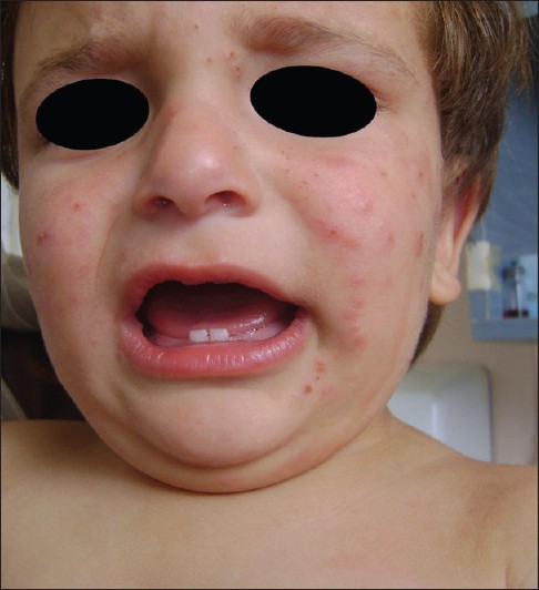
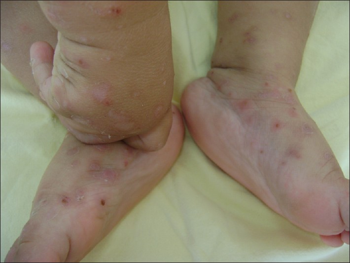
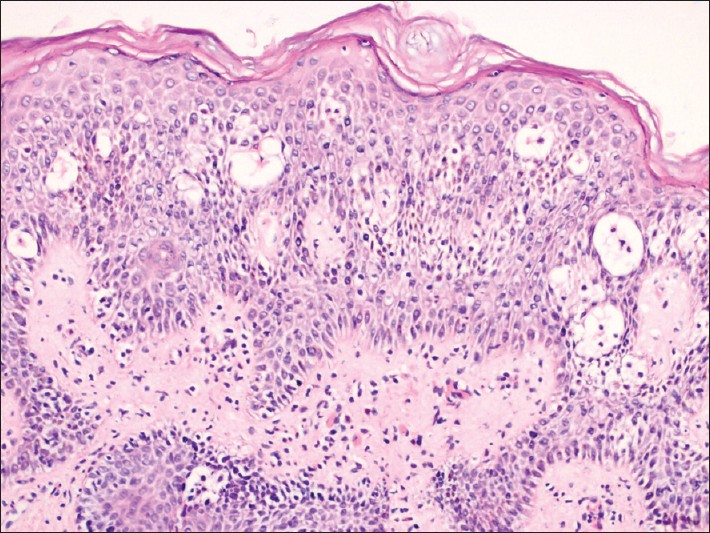
Dermatological examination revealed a papulovesicular eruption on the face and extremities and hypopigmented scars on the dorsal aspect of his hands [Figure - 1] and [Figure - 2]. The skin type of the patient was type II. The complete blood cell count and the liver and renal function tests were found to be in the normal ranges. The direct microscopic examination for Sarcoptes scabiei was negative. Histopathological examination of skin biopsy showed superficial orthokeratosis, spongiosis, irregular acanthosis, epidermal degeneration, and minimal perivascular chronic inflammatory cell infiltration in the upper dermis [Figure - 3].
 |
| Figure 1: Papulovesicular eruption on the face |
 |
| Figure 2: Papulovesicular eruption and hypopigmented scars on the hands and feet |
 |
| Figure 3: Superficial orthokeratosis, spongiosis, irregular acanthosis, epidermal degeneration, and minimal perivascular chronic inflammatory cell infiltration in the upper dermis (H and E, ×100) |
What is your Diagnosis?
| 1. |
Yesudian PD, Sharpe GR. Hydroa vacciniforme with oral mucosal involvement. Pediatr Dermatol 2004;21:555-7.
[Google Scholar]
|
| 2. |
Gupta G, Mohamed M, Kemmett D. Familial hydroa vacciniforme. Br J Dermatol 1999;140:124-6.
[Google Scholar]
|
| 3. |
Hawk JL, Young AR, Ferguson J. Cutaneous Photobiology. In: Burns T, Breathnach S, Cox N, Griffiths C, editors. Rook's Textbook of Dermatology, 7 th ed. Oxford: Blackwell Publishing; 2004. p. 1-24.
th ed. Oxford: Blackwell Publishing; 2004. p. 1-24.'>[Google Scholar]
|
| 4. |
Lysell J, Edström WD, Linde A, Carlsson G, Svennilson MJ, Westermark A, et al. Antiviral therapy in children with hydroa vacciniforme. Acta Derm Venereol 2009;89:393-7.
[Google Scholar]
|
| 5. |
Sebastian QL, Rosario RD. Hydroa vacciniforme: Differential diagnoses and workup. eMedicine Specialties, Dermatology, Photo-Related Diseases. Updated: Jul 24, 2008.
[Google Scholar]
|
| 6. |
Park HY, Park JH, Lee KT, Lee DY, Lee JH, Lee ES, et al. A case of hydroa vacciniforme. Ann Dermatol 2010;22:312-5.
[Google Scholar]
|
Fulltext Views
3,008
PDF downloads
3,226


