Translate this page into:
Pegylated liposomal doxorubicin-induced miliaria crystallina and lichenoid follicular eruption
Correspondence Address:
Wei-Sheng Chong
National Skin Centre, 1 Mandalay Road 308205
Singapore
| How to cite this article: Seghers AC, Tey HL, Tee SI, Cao T, Chong WS. Pegylated liposomal doxorubicin-induced miliaria crystallina and lichenoid follicular eruption. Indian J Dermatol Venereol Leprol 2018;84:121 |
Sir,
A 24-year-old woman was being treated for stage IV primary peritoneal carcinoma with pegylated liposomal doxorubicin at a dose of 40 mg/m2. Five days after the second dose, she developed a mildly pruritic eruption on the trunk, which became more extensive over the next 2 weeks [Figure 1a]. On inspection, her back, axillae and breasts were studded with tiny, closely spaced, clear vesicles with a silvery, shiny surface, measuring 1–3 mm in diameter [Figure 1b]. Dermoscopic examination revealed multiple translucent globules with a thin dark rim [Figure - 2]. Concurrently, there was another morphologically different eruption consisting of extensive hyperpigmented follicular keratotic papules on the trunk, neck and thighs [Figure 1a] and [Figure 1b]. The above lesions were further characterized using high-definition optical coherence tomography imaging (Skintell®, Agfa HealthCare, Belgium) and the vesicles and hyperkeratotic papules were found to be intraepidermal in nature [Figure - 3]. In addition to these lesions, the patient also developed palmar erythema and superficial erosions in the intertriginous areas [Figure 1a]. The patient's body mass index was 30 kg/m2.
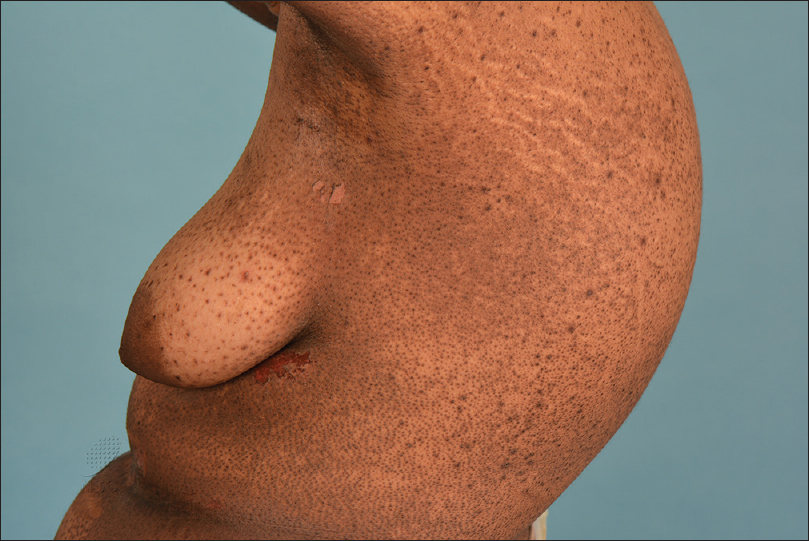 |
| Figure 1a: Extensive follicular rash affecting the trunk and erosions affecting the intertriginous areas of a young woman were seen clinically 5 days after the administration of the second dose of pegylated liposomal doxorubicin |
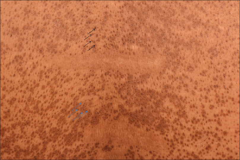 |
| Figure 1b: On closer inspection, a striking follicular eruption (blue arrow) in combination with miliaria crystallina (black arrow) were noted |
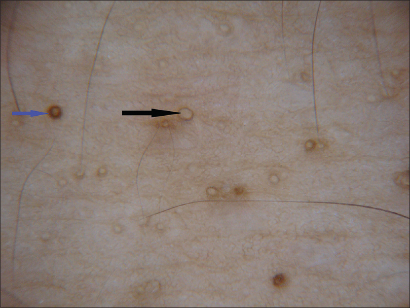 |
| Figure 2: Dermoscopy revealed multiple translucent globules with a thin dark rim; these were miliaria crystallina (black arrow). There were also round lesions with a darker and broader rim; these were the keratotic follicular papules (blue arrow). Dermlite II Pro in cross-polarisation mode; x10 magnification |
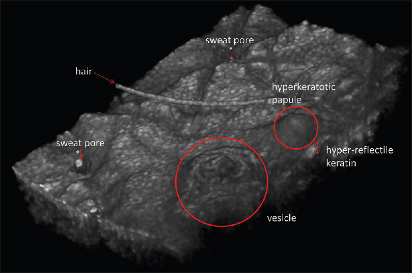 |
| Figure 3: Three-dimensional reconstruction of high-definition optical coherence tomography images demonstrating the surface topography of a vesicle and a hyperkeratotic papule. On cross-sectional view, these lesions were intraepidermal in nature and hyperrefractile keratin was apparent in the epidermis |
A skin biopsy from a vesicle on the trunk [Figure 4a] revealed the location of vesicle to be within the stratum corneum, enclosed by a thin layer of keratin. The lesion was directly overlying an acrosyringium although the associated eccrine duct was unaffected. There was minimal spongiosis or inflammation within the surrounding dermis. A separate biopsy performed from a follicular papule [Figure 4b] showed a large keratin plug protruding outwards from the infundibulum. The follicular epithelium showed lichenoid changes comprising basal vacuolar alteration and scattered necrotic keratinocytes. A sparse, superficial perivascular lymphocytic infiltrate was also present.
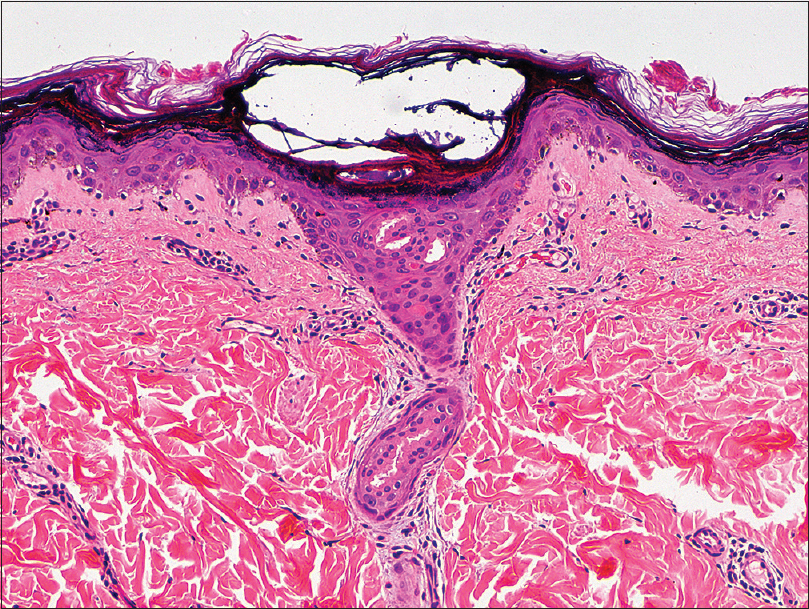 |
| Figure 4a: An intracorneal vesicle was present atop an acrosyringium, consistent with the clinical finding of miliaria crystallina (H and E, ×100) |
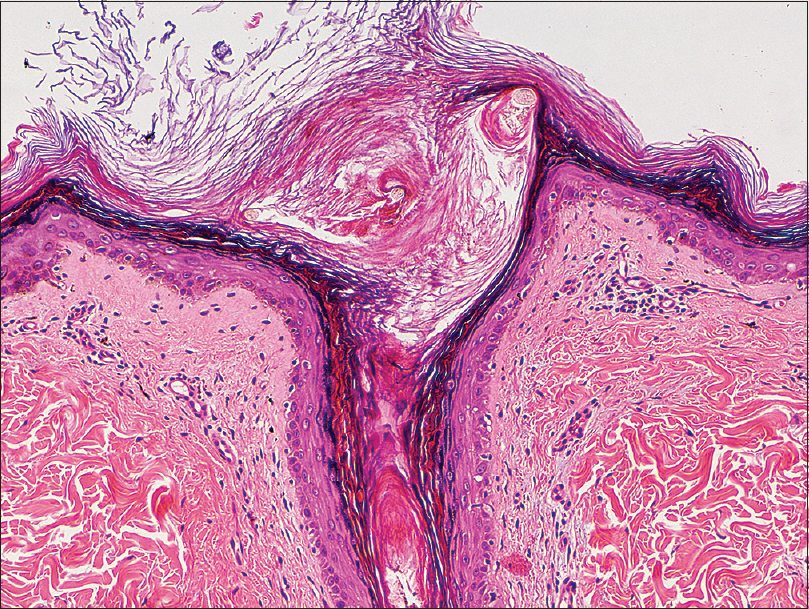 |
| Figure 4b: A keratin plug filled a dilated infundibulum and the follicular epithelium showed subtle lichenoid change comprising vacuolar alteration and keratinocyte necrosis (H and E, ×100) |
The histopathological findings were consistent with miliaria crystallina in the first specimen and a lichenoid follicular eruption in the second specimen while the clinical finding of acral erythema was in keeping with the description of palmoplantar erythrodysesthesia. Given the temporal sequence, these symptoms were consistent with an adverse reaction to pegylated liposomal doxorubicin. In view of the limited disease morbidity, she persisted with her chemotherapy, while her skin was treated with emollients and potent topical steroids. The rash remained persistent 2 months after onset.
Pegylated liposomal doxorubicin is approved for the treatment of multiple malignancies. It is a formulation of doxorubicin in which doxorubicin hydrochloride is encapsulated within pegylated liposomes.[1] This changes the biodistribution and prolongs the half-life of the drug. The liposomal formulation also reduces the concentration of free doxorubicin in the plasma and allows for greater tolerability due to fewer toxic effects, such as cardiotoxicity.[1] However, liposomal formulations deposit a substantial fraction of the drug in the skin and this may lead to mucocutaneous adverse reactions. Pegylated liposomal doxorubicin has been observed in the sweat, present within the excretory ducts of the eccrine glands and around the acrosyringium.[2] It is postulated that sweat functions as a carrier of doxorubicin to the skin surface. This is presumably made possible by the hydrophilic coating of the liposomes. After excretion onto the skin surface, the stratum corneum becomes covered with doxorubicin-enriched sweat, which may be able to penetrate into the exposed skin.
A well-known cutaneous side effect associated with pegylated liposomal doxorubicin is palmoplantar erythrodysesthesia (also known as toxic acral erythema or hand-foot syndrome) which is dose-dependent and occurs in up to 50% of patients receiving pegylated liposomal doxorubicin at 50 mg/m2 every 4 weeks.[3] This and several other overlapping clinical entities have been grouped together and termed toxic erythema of chemotherapy on the basis of shared histopathological features including interface dermatitis and dyskeratosis. This is suggestive of a common pathophysiology which, most likely, involves direct drug toxicity to the cells of the eccrine glands and the hair follicles.[4] The different skin sites involved are those areas with multiple eccrine glands and those with increased friction/warmth resulting in hyperhidrosis. Less attention is given to a follicular eruption which has been reported in the literature as diffuse follicular rash or folliculo-centric lichenoid dermatitis.[5] Another rarely reported adverse effect is miliaria crystallina which was reported in a woman 5 days after initiating pegylated liposomal doxorubicin for multiple myeloma.[6]
In our case, the patient experienced palmoplantar erythrodysesthesia following administration of doxorubicin which falls within the spectrum of toxic erythema of chemotherapy. The time of onset was also consistent, occurring between 2 days and 3 weeks of drug usage.[4] She simultaneously developed widespread miliaria crystallina and a lichenoid follicular eruption. While neither of these conditions is classically described with toxic erythema of chemotherapy, it is probable that all are incited through the same pathomechanism, ie-doxorubicin elimination via the eccrine glands inducing local necrosis and causing blockade at the acrosyringium, leading to the formation of miliaria. Doxorubicin also causes toxic damage to the follicular basal epithelium, inducing vacuolar change and dyskeratosis with a resultant lichenoid follicular eruption. Our patient had a high body mass index which might explain toxic erythema affecting the intertriginous regions in addition to the palms and soles.
Toxic erythema of chemotherapy is characterized by burning pain and redness affecting various parts of the body secondary to a dose-dependent administration of chemotherapeutic drugs which is probably due to damage to the cutaneous cells. Currently described entities affect hands and feet, intertriginous zones and less often, the elbows, knees and ears.[4] So far, no entities have been described involving other parts of the body. Frequently, these toxic reactions are misdiagnosed as hypersensitivity reactions as well as graft-versus-host disease, vasculitis or cutaneous infections. However, the condition is self-limited and resolves with desquamation and post-inflammatory hyperpigmentation.
In conclusion, we describe a patient who, apart from experiencing classical palmoplantar erythrodysesthesia and intertrigo-like eruption after doxorubicin, simultaneously also developed miliaria crystallina and a diffuse lichenoid follicular eruption. We propose that these reactions be considered as part of the spectrum of clinical features encountered in toxic erythema of chemotherapy. Increased awareness of this condition will result in more accurate diagnosis and better management of the patients, including reassurance and cooling techniques.
Financial support and sponsorship
Nil.
Conflicts of interest
There are no conflicts of interest.
| 1. |
Gabizon A, Martin F. Polyethylene glycol-coated (pegylated) liposomal doxorubicin. Rationale for use in solid tumours. Drugs 1997;54 Suppl 4:15-21.
[Google Scholar]
|
| 2. |
Jacobi U, Waibler E, Schulze P, Sehouli J, Oskay-Ozcelik G, Schmook T, et al. Release of doxorubicin in sweat:First step to induce the palmar-plantar erythrodysesthesia syndrome? Ann Oncol 2005;16:1210-1.
[Google Scholar]
|
| 3. |
Lorusso D, Di Stefano A, Carone V, Fagotti A, Pisconti S, Scambia G. Pegylated liposomal doxorubicin-related palmar-plantar erythrodysesthesia ('hand-foot' syndrome). Ann Oncol 2007;18:1159-64.
[Google Scholar]
|
| 4. |
Bolognia JL, Cooper DL, Glusac EJ. Toxic erythema of chemotherapy: A useful clinical term. J Am Acad Dermatol 2008;59:524-9.
[Google Scholar]
|
| 5. |
Dai J, Micheletti R, Rosenbach M, Chu EY. Striking follicular eruption to pegylated liposomal doxorubicin. Am J Dermatopathol 2014;36:590-1.
[Google Scholar]
|
| 6. |
Godkar D, Razaq M, Fernandez G. Rare skin disorder complicating doxorubicin therapy: Miliaria crystallina. Am J Ther 2005;12:275-6.
[Google Scholar]
|
Fulltext Views
3,480
PDF downloads
3,386


