Translate this page into:
Post-traumatic ectopic nail
2 Department of Pathology, North Eastern Indira Gandhi Regional Institute of Health and Medical Sciences, Shillong, Meghalaya, India
Correspondence Address:
Shikha Verma
Department of Dermatology and STD, North Eastern Indira Gandhi Regional Institute of Health and Medical Sciences, Shillong, Meghalaya
India
| How to cite this article: Thakur BK, Verma S, Jitani A. Post-traumatic ectopic nail. Indian J Dermatol Venereol Leprol 2016;82:416 |
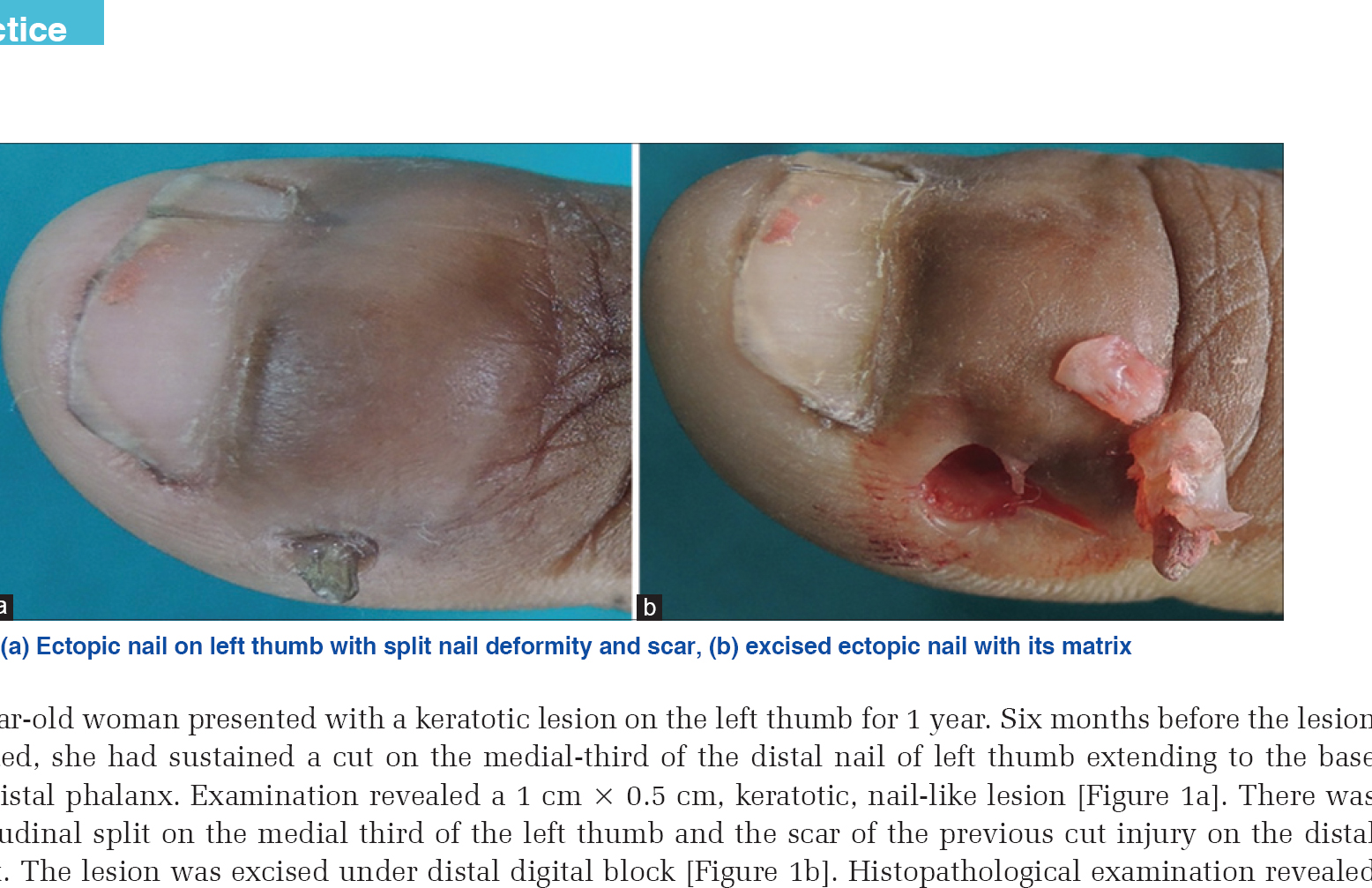
A 38-year-old woman presented with a keratotic lesion on the left thumb for 1 year. Six months before the lesion was noted, she had sustained a cut on the medial-third of the distal nail of left thumb extending to the base of the distal phalanx. Examination revealed a 1 cm × 0.5 cm, keratotic, nail-like lesion [Figure - 1]a. There was a longitudinal split on the medial third of the left thumb and the scar of the previous cut injury on the distal phalanx. The lesion was excised under distal digital block [Figure - 1]b. Histopathological examination revealed nail plate and nail matrix confirming the diagnosis of ectopic nail.
 |
| Figure 1: (a) Ectopic nail on left thumb with split nail deformity and scar, (b) excised ectopic nail with its matrix |
Financial support and sponsorship
Nil.
Conflicts of interest
There are no conflicts of interest.
Fulltext Views
3,351
PDF downloads
3,235


