Translate this page into:
Radiofrequency-assisted fractional thermolysis for drug delivery in Keloids
Corresponding author: Prof. Somesh Gupta, Department of Dermatology and Venereology, All India Institute of Medical Sciences, New Delhi, Delhi, India. someshgupta@aiims.edu
-
Received: ,
Accepted: ,
How to cite this article: Choudhary R, Bharti P, Mondal A, Gupta S. Radiofrequency-assisted fractional thermolysis for drug delivery in Keloids. Indian J Dermatol Venereol Leprol. 2024;90:842-3. doi: 10.25259/IJDVL_647_2023
Problem
Trans-epidermal large molecule drug delivery is challenging. Fractional ablative lasers, dermabrasion and micro-needling have been used for this purpose.1 As these procedures create channels only up to the superficial dermis, delivery of drugs into thick keloids is inadequate and it often leads to an unsatisfactory response.2 Apart from this, devices like fractional lasers are not available at all dermatology centres. Intralesional radiofrequency ablation has been used for assisted drug deposition in keloids; however, this does not lead to uniform dispersion of the drug.3
Solution
We describe a novel technique of radiofrequency-assisted fractional thermolysis that creates three-dimensional deep channels which can be customised according to the thickness of the keloid. After administering local anaesthesia, markings for points of entry were made at regular intervals [Video 1]. A pointed radiofrequency probe (KLS Martin, Tuttligen, Germany), set at 20-Watt power in coagulation mode, was used to create full-thickness pores [Video 1]. In thicker lesions, pores were also created horizontally from the sides of the lesions, making it a three-dimensional process. Triamcinolone acetonide (40 mg/mL) was gently massaged over the lesion and the lesion was covered with an occlusive film dressing for 24 h. Patients continued this application at home for the next 5–7 days. The technique is based on direct collagen remodelling along with drug delivery to the full depth of the lesion. It causes fractional destruction of the tissue till the full depth of the lesion, leading to penetration of the drug till the full depth and faster healing as compared to superficial shaving of the lesion. This technique was used in eight patients with recalcitrant pre-sternal keloids. After three sessions, the resolution of symptoms (itching and pain) was seen in all patients. The mean patient-perceived improvement was 80.62 ± 9.42%, whereas the mean physician-perceived reduction in size was 63.12 ± 11.63% [Figure 1a, 1b, 1c, 2a and 2b]. Secondary infection was noted in one patient, which resolved with oral antibiotics. None of the patients developed atrophy or dyspigmentation. It is a novel technique introduced by authors and is yet to be compared with the standard approach. The technique was tried in patients who had previously failed conventional treatment including intralesional injections. Further studies with a larger sample size and longer follow-up periods are needed to confirm the efficacy and safety of this technique. Apart from triamcinolone acetonide, this technique can also be combined with topical hyaluronidase, 5-fluorouracil, verapamil, and bleomycin.
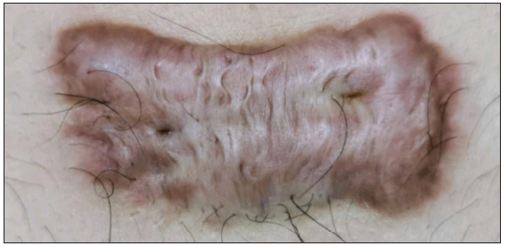
- Baseline image of keloid over chest.
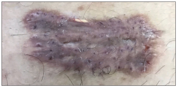
- Flattening of keloid after 2 sessions.
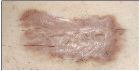
- Significant flattening of keloid after three sessions and 1 month follow up.
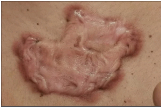
- Baseline photo of keloid over chest.
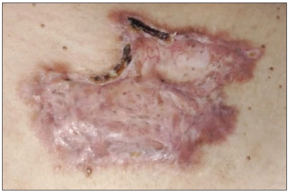
- Significant improvement after 3 sessions.
Declaration of patient consent
The authors certify that they have obtained all appropriate patient consent.
Financial support and sponsorship
Nil.
Conflicts of interest
There are no conflicts of interest.
Use of artificial intelligence (AI)-assisted technology for manuscript preparation
The authors confirm that there was no use of artificial intelligence (AI)-assisted technology for assisting in the writing or editing of the manuscript and no images were manipulated using AI.
References
- Laser-assisted drug delivery. Actas Dermo-Sifiliográficas (English Edition). 2018;109:858-67.
- [CrossRef] [PubMed] [Google Scholar]
- A review of current keloid management: mainstay monotherapies and emerging approaches. Dermatol Ther (Heidelb). 2020;10:931-48.
- [CrossRef] [PubMed] [PubMed Central] [Google Scholar]
- Modified technique of intralesional radiofrequency with drug deposition in keloids using customized intravenous cannulas. J Am Acad Dermatol. 2021;89(2):e89-e90.
- [CrossRef] [PubMed] [Google Scholar]


