Translate this page into:
Reflectance confocal microscopy characteristics of trichofolliculoma
Corresponding author: Dr. HongYong Sun, Department of Dermatology, Inner Mongolia Autonomous Region People’s Hospital, Hohhot, Inner Mongolia Autonomous Region, China. 418217546@qq.com
-
Received: ,
Accepted: ,
How to cite this article: Sun HY, Wang J, Liu F, Li R. Reflectance confocal microscopy characteristics of trichofolliculoma. Indian J Dermatol Venereol Leprol. 2025;91:100-3. doi: 10.25259/IJDVL_714_2023
Dear Editor,
Trichofolliculoma is a relatively rare hair matrix malformation tumour, which can be diagnosed primarily on histopathological examination. Dermoscopic features of trichofolliculoma have been infrequently reported, and only one study has described its reflectance confocal microscopy (RCM) characteristics.1 Thus, there is no consensus on the characteristics of RCM for trichofolliculoma, and the use of this modality for trichofolliculoma remains in the exploratory stage. We report the RCM findings of a lesion histologically diagnosed as trichofolliculoma.
A 44-year-old man presented with a three-year history of an asymptomatic hemispherical papule on the right side of his nose. The light red hemispherical papule which was located on the medial aspect of right ala measured approximately 0.4 cm x 0.5 cm. The lesion had a smooth surface and a central depression filled with a yellowish-white, keratin-like material, and was not tender. There were no hair structures visible on the surface to the naked eye [Figure 1a]. Dermoscopic examination revealed nonspecific features: pinkish-white, uniform, structureless areas with two central yellowish-white areas,2 protruding black hair, brownish spots, and peripheral radiating bright white streaks accompanied with linear blood vessels [Figure 1b]. Previously reported patterns, such as ‘mouldy peach’, ‘troll doll hair’, and ‘fireworks’, were not observed.3–5
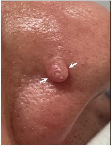
- A light red hemispherical papule with a smooth surface and a central depression filled with yellowish-white, keratin-like material (white arrow).
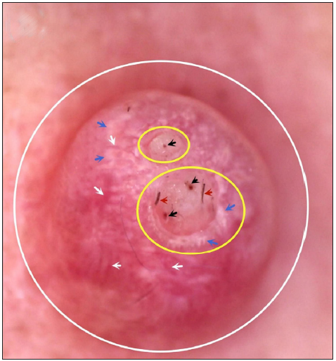
- Dermoscopy: A uniform, unstructured pink and pinkish-white hemispherical papule (white circle) with two central yellowish-white depressions (yellow circle), protruding black hair (red arrow), brownish spots (black arrow), and radiating peripheral bright white streaks (blue arrow) accompanied by linear blood vessels (white arrow).
RCM examination of the patient’s lesion revealed the following characteristics: 1) thinned epidermis with focal low-refractive areas and a visible normal honeycomb structure [Figure 2a]; 2) a reduced number of dermal papillary rings, with some appearing as semicircular crescents and with increased spacing (previously unreported) [Figure 2b]; 3) expanded hair follicles in the superficial and middle dermis, exhibiting medium-to-low refraction, and plump finger-like protrusions (medium to high refraction) extending along the hair follicles. Overall, the morphology resembled a ‘rabbit’, with flattened oval and elongated clumps of varying sizes radiating around the expanded hair follicles, appearing as ‘cloud-like radial flow’[Figure 2c]. 4) uniform-sized clumps resembling cobblestones and exhibiting medium-to-high refraction are seen in Figure 2d; 5) thickened collagen fibres are arranged in bundles, adjacent to hair follicles and within the dermis [Figure 2e].
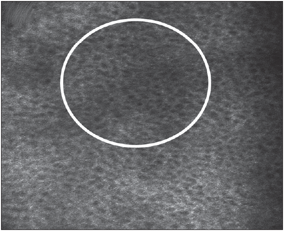
- Focal thinning of the epidermis visible as low-refractive dark areas (white circle).
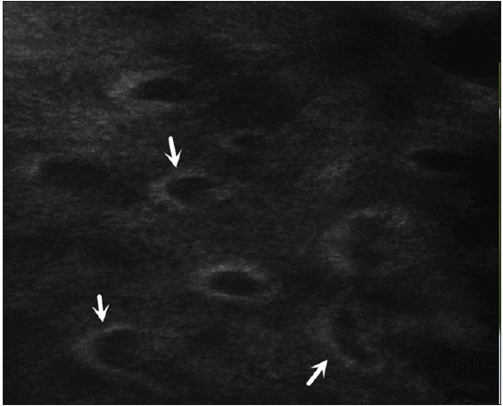
- A decreased number of dermal papillary rings, with some appearing as semi-circular and having increased spacing (white arrows).
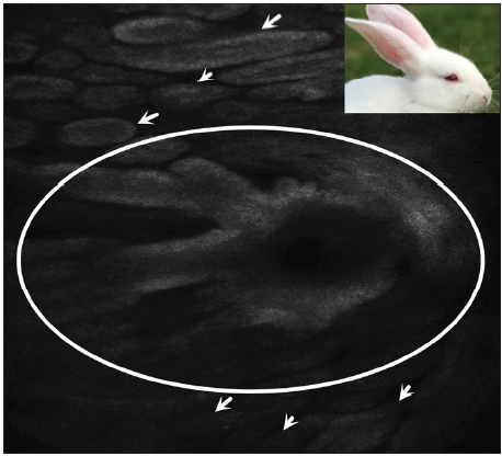
- Expanded hair follicles in the superficial and middle dermis exhibiting medium-to-low refraction, and plump finger-like protrusions extending along the hair follicles with medium-to-high refraction (white circles). Variably sized flattened oval and elongated clumps radiating around the expanded hair follicles, presenting a ‘cloud-like radial flow’ appearance (white arrows), rabbit like pattern (inset).
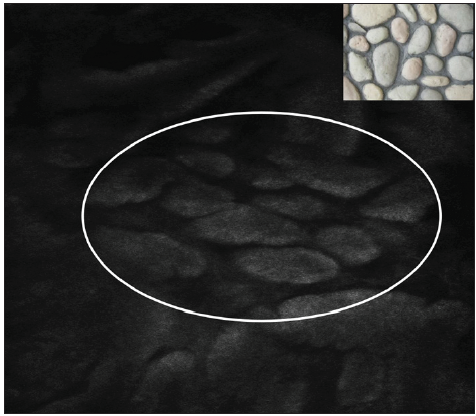
- Clumps in certain areas that are relatively uniform in size (white circle), resembling a cobblestone-like appearance (inset).
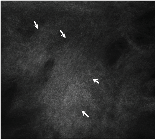
- Bundled, thickened collagen fibres adjacent to hair follicles and within the dermis (white arrows).
Histology demonstrated a prominent expansion of hair follicles in the dermis, containing keratinous material within the follicles. The follicular epithelium extended peripherally in cord-like formations, giving rise to numerous secondary follicular cavities in a radiating distribution, including secondary or tertiary follicles as seen in Figure 2f. Based on the histopathological features, a diagnosis of trichofolliculoma was confirmed.
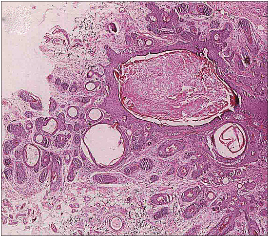
- Pronounced expansion of hair follicles in the dermis, containing keratinous material within the follicles and epithelium extending peripherally in cord-like formations, generating numerous secondary follicular cavities in a radiating distribution and secondary or tertiary follicles (Haematoxylin and eosin, 100x).
The findings in our case are mostly consistent with those described by Karaarslan et al,1 which could allow a consensus on diagnostic findings for this entity.1 Our attempt to use metaphorical terms such as ‘rabbit sign, cloud sign, radial flow’, could help clinical dermatologists better grasp the reflectance confocal microscope features of trichofolliculoma. More importantly, there are some newly discovered microscopic features, such as the ‘cobblestone sign’, which still needs further corroboration. Furthermore, by comparing with histopathological images, we found that there might be a correlation between the partial RCM features of trichofolliculoma and its histopathological features. For example, there might be a corresponding relationship between the ‘rabbit sign’ appearing under a reflectance confocal microscope and the dilated hair follicles and their elongated wall epithelium in histopathology, while a large number of secondary and tertiary hair follicle cavities derived from around the central hair follicle in histopathology might correspond to the cloud sign, cobblestone sign, and radial flow structures appearing under a reflectance confocal microscope. However, being a single case analysis, this is merely speculative and requires further study on a larger number of cases.
We have hereby described the RCM characteristics of trichofolliculoma. RCM could emerge as a pivotal diagnostic tool in cases with non-diagnostic dermoscopic findings.
Declaration of patient consent
The authors certify that they have obtained all appropriate patient consent.
Financial support and sponsorship
Nil.
Conflicts of interest
There are no conflicts of interest
Use of artificial intelligence (AI)-assisted technology for manuscript preparation
The authors confirm that there was no use of artificial intelligence (AI)-assisted technology for assisting in the writing or editing of the manuscript and no images were manipulated using AI.
Reference
- In vivo reflectance confocal microscopic findings in a case of trichofolliculoma. Anais Brasileiros de Dermatologia. 2022;97:236-9.
- [CrossRef] [PubMed] [Google Scholar]
- Trichofolliculoma: A retrospective review of 8 cases. Ann. Dermatol. Venereol. 2015;142:183-8.
- [CrossRef] [PubMed] [Google Scholar]
- The “firework” pattern in dermoscopy. Int J Dermatol. 2013;52:1158-9.
- [CrossRef] [PubMed] [Google Scholar]
- Dermoscopy of trichofolliculoma: A rare hair follicle hamartoma. J Eur Acad Dermatol Venereol. 2017;31:e123-e124.
- [CrossRef] [PubMed] [Google Scholar]
- ‘Mouldy peach’ dermoscopic pattern in trichofolliculoma. J EurAcad Dermatol Venereol. 2022;36:e726-e727.
- [Google Scholar]


