Translate this page into:
Scrotal plaques as a predominant presentation in a case of secondary syphilis
Corresponding author: Dr. Anupama Bains, Department of Dermatology, AIIMS, First Floor, OPD Building, Basni Phase 2, Jodhpur, Rajasthan, India. whiteangel2387@gmail.com
-
Received: ,
Accepted: ,
How to cite this article: Bains A, Tyagi N. Scrotal plaques as a predominant presentation in a case of secondary syphilis. Indian J Dermatol Venereol Leprol 2021;87:252-4.
Sir,
A 28-year-old male presented to the dermatology out-patient department with a 20-day history of multiple, mildly pruritic, erythematous lesions over the scrotum. There was a history of unprotected sexual exposure with a commercial sex worker in the 3 months back. General physical examination and systemic examination revealed no significant abnormalities. Cutaneous examination showed multiple, erythematous, flat-topped, round to oval, firm, non-tender plaques distributed over the scrotum [Figure 1a]. A few pigmented macules were seen over the soles [Figure 1b]. Mucosal examination revealed no abnormal findings. A skin biopsy specimen from a lesion over the scrotum revealed perivascular lymphohistiocytic inflammation admixed with numerous plasma cells [Figure 2a and b]. A serology test revealed a reactive Venereal Disease Research Laboratory test with a titer of 1:32 and a positive Treponema pallidum hemagglutinin assay. Patient was treated with benzathine penicillin 2.4 million units IM which led to complete resolution of lesions at 4 weeks [Figure 2c].
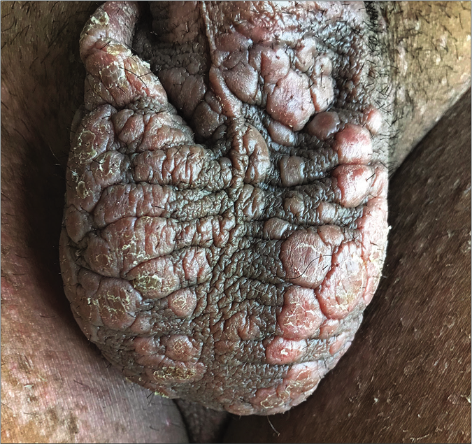
- Multiple erythematous, flat-topped, round to oval plaques over the scrotum
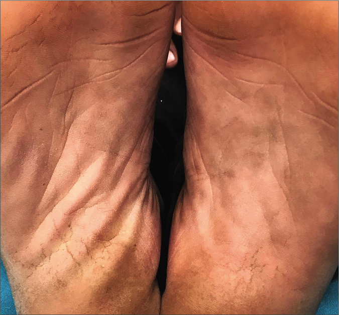
- A few pigmented macules over instep of soles
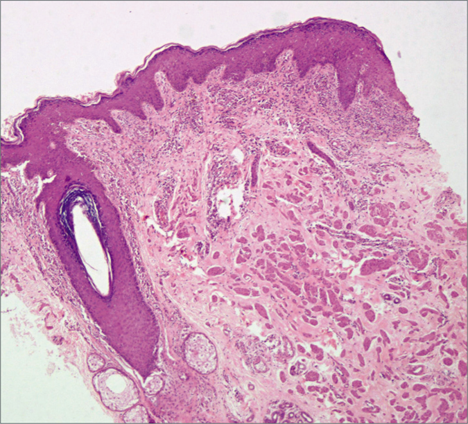
- Histopathology shows acanthosis along with perivascular chronic mononuclear inflammatory cells. (H&E, 4×)
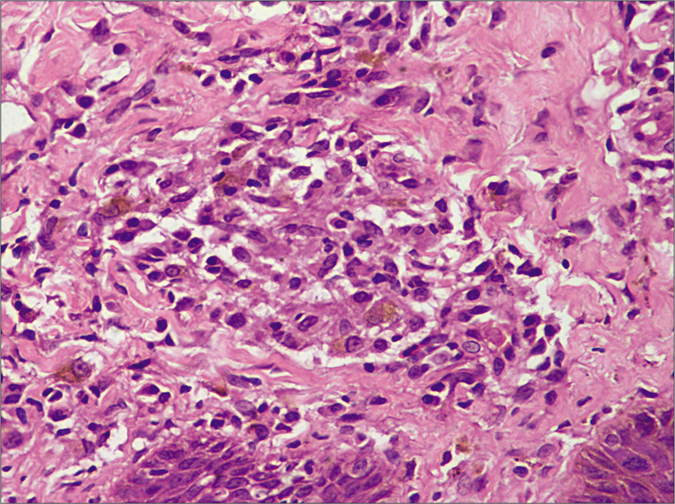
- Higher magnification shows loose collection of histiocytes admixed with plasma cells in the dermis (H&E, 40×)
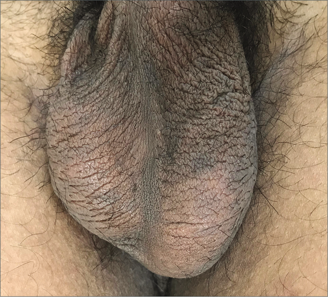
- Resolution of scrotal lesions after treatment
Secondary syphilis is often called the “great imitator” as it can have a variety of clinical presentations. Lichenoid plaques on the scrotum and scrotal dermatitis have been rarely described in literature.1-6 This case was interesting as he had no generalized rash, mucosal lesions or other systemic manifestations of secondary syphilis. Cases described with scrotal lesions had predominantly lichenoid morphology along with the presence of other features of secondary syphilis.1,5 A few patients had erythematous scaly plaques which were confused with eczema. Onset of the scrotal lesions preceded condyloma lata by several months in another patient.3 In our case too, the scrotal lesions were the chief complaints and could be an early manifestation of secondary syphilis. However, further studies are required to conclude this finding. A few slightly pigmented asymptomatic macules over the palms and soles may remain unnoticed by the patient, especially in Indian skin. Hence, syphilis should be considered as an important differential diagnosis in patients presenting with scrotal lesions alone as it can mimic other dermatoses like lichen planus, psoriasis and eczema.
Declaration of patient consent
The authors certify that they have obtained all appropriate patient consent.
Financial support and sponsorship
Nil.
Conflicts of interest
There are no conflicts of interest.
References
- Penile edema and lichenoid plaques on scrotum: An unusual presentation of secondary syphilis. Indian Dermatol Online J. 2019;10:585-6.
- [CrossRef] [PubMed] [Google Scholar]
- Secondary syphilis: An unusual presentation. Indian J Sex Transm Dis AIDS. 2017;38:98-9.
- [CrossRef] [PubMed] [Google Scholar]
- Secondary syphilis presenting as scrotal eczema. J Am Acad Dermatol. 2007;57:1099-101.
- [CrossRef] [PubMed] [Google Scholar]
- Flesh colored plaques on the scrotum. J Am Acad Dermatol. 2015;72:e75-6.
- [CrossRef] [Google Scholar]
- Secondary syphilis presenting as annular lichenoid plaques on the scrotum. J Cutan Med Surg. 2008;12:114-6.
- [CrossRef] [PubMed] [Google Scholar]
- Lichenoid skin lesions: A rare manifestation in secondary syphilis. Indian J Dermatol Venereol Leprol. 2020;86:105.
- [CrossRef] [PubMed] [Google Scholar]


