Generic selectors
Exact matches only
Search in title
Search in content
Post Type Selectors
Search in posts
Search in pages
Filter by Categories
15th National Conference of the IAOMFP, Chennai, 2006
Abstract
Abstracts from current literature
Acne in India: Guidelines for management - IAA Consensus Document
Addendum
Announcement
Art & Psychiatry
Article
Articles
Association Activities
Association Notes
Award Article
Book Review
Brief Report
Case Analysis
Case Letter
Case Letters
Case Notes
Case Report
Case Reports
Clinical and Laboratory Investigations
Clinical Article
Clinical Studies
Clinical Study
Commentary
Conference Oration
Conference Summary
Continuing Medical Education
Correspondence
Corrigendum
Cosmetic Dermatology
Cosmetology
Current Best Evidence
Current Issue
Current View
Derma Quest
Dermato Surgery
Dermatopathology
Dermatosurgery Specials
Dispensing Pearl
Do you know?
Drug Dialogues
e-IJDVL
Editor Speaks
Editorial
Editorial Remarks
Editorial Report
Editorial Report - 2007
Editorial report for 2004-2005
Errata
Erratum
Focus
Fourth All India Conference Programme
From Our Book Shelf
From the Desk of Chief Editor
General
Get Set for Net
Get set for the net
Guest Article
Guest Editorial
History
How I Manage?
IADVL Announcement
IADVL Announcements
IJDVL Awards
IJDVL AWARDS 2015
IJDVL Awards 2018
IJDVL Awards 2019
IJDVL Awards 2020
IJDVL International Awards 2018
Images in Clinical Practice
Images in Dermatology
In Memorium
Inaugural Address
Index
Knowledge From World Contemporaries
Leprosy Section
Letter in Response to Previous Publication
Letter to Editor
Letter to the Editor
Letter to the Editor - Case Letter
Letter to the Editor - Letter in Response to Published Article
LETTER TO THE EDITOR - LETTERS IN RESPONSE TO PUBLISHED ARTICLES
Letter to the Editor - Observation Letter
Letter to the Editor - Study Letter
Letter to the Editor - Therapy Letter
Letter to the Editor: Articles in Response to Previously Published Articles
Letters in Response to Previous Publication
Letters to the Editor
Letters to the Editor - Letter in Response to Previously Published Articles
Letters to the Editor: Case Letters
Letters to the Editor: Letters in Response to Previously Published Articles
Media and news
Medicolegal Window
Messages
Miscellaneous Letter
Musings
Net Case
Net case report
Net Image
Net Images
Net Letter
Net Quiz
Net Study
New Preparations
News
News & Views
Obituary
Observation Letter
Observation Letters
Oration
Original Article
ORIGINAL CONTRIBUTION
Original Contributions
Pattern of Skin Diseases
Pearls
Pediatric Dermatology
Pediatric Rounds
Perspective
Presedential Address
Presidential Address
Presidents Remarks
Quiz
Recommendations
Regret
Report
Report of chief editor
Report of Hon : Treasurer IADVL
Report of Hon. General Secretary IADVL
Research Methdology
Research Methodology
Resident page
Resident's Page
Resident’s Page
Residents' Corner
Residents' Corner
Residents' Page
Retraction
Review
Review Article
Review Articles
Reviewers 2022
Revision Corner
Self Assessment Programme
SEMINAR
Seminar: Chronic Arsenicosis in India
Seminar: HIV Infection
Short Communication
Short Communications
Short Report
Snippets
Special Article
Specialty Interface
Studies
Study Letter
Study Letters
Supplement-Photoprotection
Supplement-Psoriasis
Symposium - Contact Dermatitis
Symposium - Lasers
Symposium - Pediatric Dermatoses
Symposium - Psoriasis
Symposium - Vesicobullous Disorders
SYMPOSIUM - VITILIGO
Symposium Aesthetic Surgery
Symposium Dermatopathology
Symposium-Hair Disorders
Symposium-Nails Part I
Symposium-Nails-Part II
Systematic Review and Meta-Analysis
Systematic Reviews and Meta-analyses
Systematic Reviews and Meta-analysis
Tables
Technology
Therapeutic Guideline-IADVL
Therapeutic Guidelines
Therapeutic Guidelines - IADVL
Therapeutics
Therapy
Therapy Letter
Therapy Letters
View Point
Viewpoint
What’s new in Dermatology
View/Download PDF

Translate this page into:

Letter to the Editor - Case Letter
2016:82:4;455-456
doi: 10.4103/0378-6323.174415
PMID: 27279319

Two cases of axillary chromoblastomycosis

Sowmyashree Krishna1 , Manjunath M Shenoy1 , Malcolm Pinto1 , Varsha Saxena2
1 Department of Dermatology, Yenepoya Medical College, Yenepoya University, Mangalore, Karnataka, India
2 Department of Microbiology, Yenepoya Medical College, Yenepoya University, Mangalore, Karnataka, India

Correspondence Address:
Manjunath M Shenoy
Department of Dermatology, Yenepoya Medical College, Yenepoya University, Deralakatte, Mangalore - 575 018, Karnataka
India
How to cite this article:
Krishna S, Shenoy MM, Pinto M, Saxena V. Two cases of axillary chromoblastomycosis. Indian J Dermatol Venereol Leprol 2016;82:455-456
Copyright: (C)2016 Indian Journal of Dermatology, Venereology, and Leprology

Sir,

Chromoblastomycosis is a chronic fungal infection of the skin and subcutaneous tissue caused by dematiaceous fungi, the most common causative organisms being Fonsecaea pedrosoi, Phialophora verrucosa, Fonsecaea compacta and Cladophialophora carionni.[1] The hallmark of chromomycosis is sclerotic bodies which can be demonstrated in potassium hydroxide mount and routine hematoxylin and eosin staining.[2] Lower limbs are the most commonly affected sites. Unusual extracutaneous sites of involvement include pleural cavity, ileocecal region, laryngotracheal area and tonsils.[3] Dissemination, though rare, presents with multiple verrucous nodules and plaques over limbs, neck, face, lymph glands and tonsil according to reports.[4] The most frequent complication is secondary bacterial infection but malignancies have also been recorded.[2]

A 54-year-old farmer with no prior history of trauma presented with an asymptomatic erythematous plaque over the right axilla for 1 year. General physical and systemic examination was unremarkable and baseline investigations were found to be within normal limits. On cutaneous examination, a solitary, well-defined erythematous plaque measuring about 6 cm × 4 cm was present over the lateral wall of right axilla [Figure - 1]. On closer examination, a few black dots were present over the surface of the lesion. There were no satellite lesions. A provisional diagnosis of lupus vulgaris and chromoblastomycosis was made.
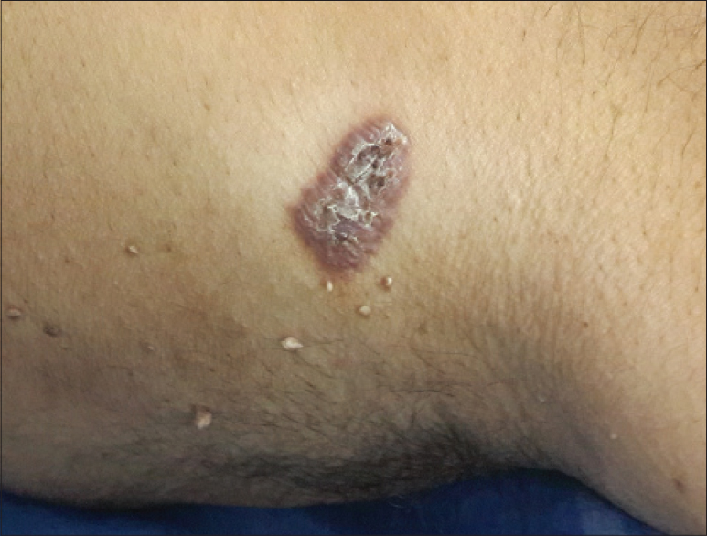
Figure 1: A single, well-defined erythematous plaque over lateral wall of right axilla

A 37-year-old cook presented with asymptomatic skin lesions over the axilla for 2 years. It started as a papule which slowly increased in size over the next 2 years. The second lesion developed after a span of 4 months. The rest of the physical examination and baseline investigations were within normal limits. On cutaneous examination, there were two well-defined polycyclic plaques; the larger lesion measuring about 9 cm × 5 cm on the lateral wall of axilla and smaller lesion with central scarring measuring about 5 cm × 7 cm on the medial wall of left axilla [Figure - 2]. No regional lymphadenopathy was detected. Lupus vulgaris, chromoblastomycosis and atypical mycobacterial infection were considered in the differential diagnosis.
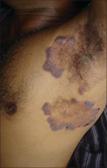
Figure 2: Two polycyclic plaques present over lateral and medial wall of left axilla

Skin scrapings with ten percent potassium hydroxide mount revealed typical brownish round thick-walled sclerotic cells in both cases. A skin biopsy was obtained from the verrucous plaque in both the cases and histopathological examination revealed hyperkeratosis, pseudoepitheliomatous hyperplasia and granulomatous inflammation with mixed cell infiltrate consisting of neutrophils, lymphocytes, epithelioid cells, multinucleated giant cells and sclerotic bodies which were diagnostic of chromoblastomycosis [Figure - 3]. Fungal culture on Sabouraud's dextrose agar yielded growth of Fonsecaea in one case and in the other case no organism associated with chromoblastomycosis could be isolated. Both were treated with itraconazole, 200 mg/day. The first patient was also administered monthly sessions of cryotherapy using liquid nitrogen. At the end of 2 months, there was noticeable improvement with reduction in size of the plaques.
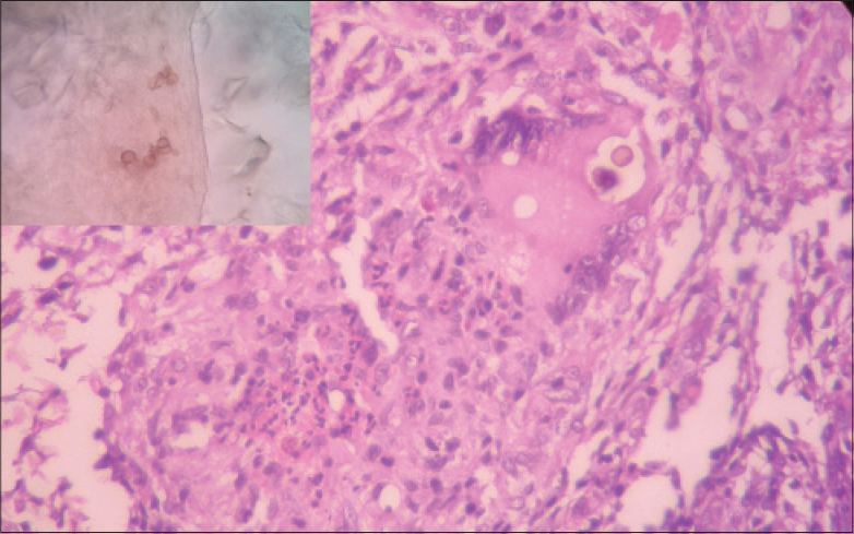
Figure 3: Skin biopsy showed mixed inflammatory cell infiltrate consisting of neutrophils, lymphocytes, epithelioid cells, multinucleated giant cells and sclerotic bodies (H and E, ×400). Inset showing sclerotic bodies on potassium hydroxide mount

We found a single previous report of axillary involvement in chromoblastomycosis in a patient who presented with genital elephantiasis due to lymphatic obstruction along with discharging sinuses and involvement of lower abdomen, face, neck, thighs and axilla.[5]

In both cases, our initial diagnosis was lupus vulgaris. Lupus vulgaris commonly affects the head and neck followed by the arms and legs. Atrophic scarring of lesions and apple jelly color on diascopy are characteristic. The clinical presentation of lupus vulgaris and chromomycosis is similar and it is difficult to differentiate between the two, especially in early cases. Despite the rarity of the site affected, potassium hydroxide mount and histopathology aided in arriving at the diagnosis of chromoblastomycosis. When chromoblastomycosis is suspected, scrapings to demonstrate Medlar bodies should be taken from an area where black dots are seen as these represent the transdermal elimination of fungal agents.[2] Potassium hydroxide mount is a simple, cost-effective bedside tool which can be employed to diagnose such cases and also useful when no facilities for fungal culture or histopathology are available.

Financial support and sponsorship

Nil.

Conflicts of interest

There are no conflicts of interest.

References
1.
López MartÍnez R, Méndez Tovar LJ. Chromoblastomycosis. Clin Dermatol 2007;25:188-94.
[Google Scholar]
2.
Ameen M. Chromoblastomycosis: Clinical presentation and management. Clin Exp Dermatol 2009;34:849-54.
[Google Scholar]
3.
Sharma NL, Sharma RC, Grover PS, Gupta ML, Sharma AK, Mahajan VK. Chromoblastomycosis in India. Int J Dermatol 1999;38:846-51.
[Google Scholar]
4.
Pavithran K. Disseminated chromoblastomycosis. Indian J Dermatol Venereol Leprol 1991;57:155-6.
[Google Scholar]
5.
Sharma N, Marfatia YS. Genital elephantiasis as a complication of chromoblastomycosis: A diagnosis overlooked. Indian J Sex Transm Dis 2009;30:43-5.
[Google Scholar]

Fulltext Views
3,057

PDF downloads
1,971
Show Sections