Translate this page into:
Two cases of D-penicillamine-induced elastosis perforans serpiginosa
Correspondence Address:
Lianjun Chen
Department of Dermatology, Fudan University, Huashan Hospital, 12 Wulumuqi Zhong Road, Jing'an District, Shanghai 200040
People�s Republic of China
| How to cite this article: Liang J, Wang D, Xu J, Chen L. Two cases of D-penicillamine-induced elastosis perforans serpiginosa. Indian J Dermatol Venereol Leprol 2016;82:452-454 |
Sir,
Elastosis perforans serpiginosa is a reactive perforating dermatosis in which elastic fibers are extruded through the epidermis. Although D-penicillamine appears to be a clear trigger for elastosis perforans serpiginosa, D-penicillamine-induced disease has been rarely reported.[1] D-penicillamine is a well-known heavy metal chelator, classically used in the treatment of Wilson's disease, rheumatoid arthritis and cystinuria. D-penicillamine-induced skin manifestations can be categorized into three groups: acute hypersensitivity reactions, bullous dermatoses and degenerative dermatoses.[2]
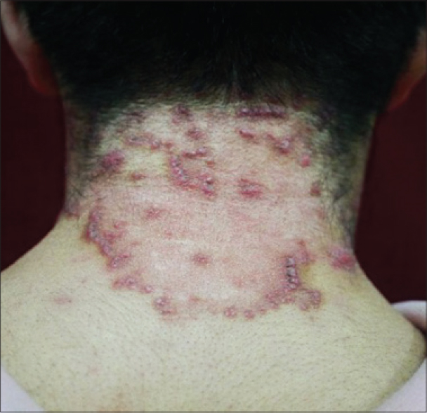
The first patient was a 32-year-old man who had been diagnosed with Wilson's disease at the age of 16 and had been taking D-Penicillamine, 0.75–1.5 g daily since then. He presented to us with a two-year history of slightly itchy, red, keratotic papules, 3–5 mm in diameter, arranged in annular, serpiginous, geographic and other irregular patterns on the neck. The annular lesions showed slight central atrophy and hyperpigmentation [Figure - 1]. He was crippled, and his eyes showed Kayser–Fleischer rings. Serum and urinary copper levels were raised on multiple occasions. A liver biopsy in 2005 had suggested cirrhosis.
 |
| Figure 1: Case 1: Red keratotic papules grouped in annular configuration on the neck |
Skin biopsy from the raised border of a lesion demonstrated a markedly acanthotic epidermis with a trans-epidermal channel containing thick, coarse elastic fibers. Abundant abnormal, clumped, elastic fibers were present in the reticular dermis. These were confirmed to be elastic fibers on staining with Verhoeff–van Gieson stain [Figure - 2]. The striking feature noted at higher magnification was the presence of multiple serrations and buds arising perpendicularly from the elastic fibers [Figure - 3]. It was not possible to discontinue D-penicillamine because no good alternative for the treatment of Wilson's disease is available in China. He was advised to use tazarotene gel topically which resulted in slow improvement.
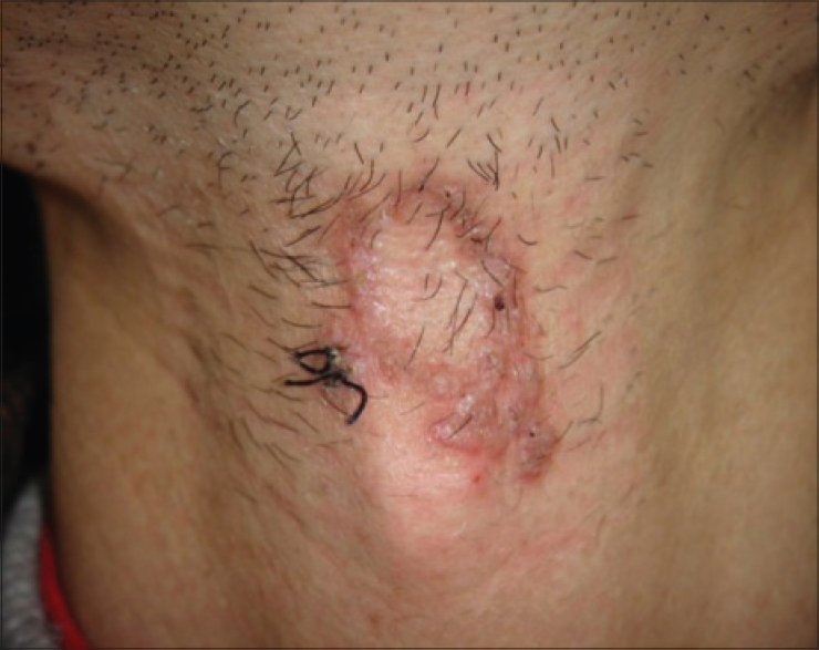
The second patient was a 38-year-old man, also a known case of Wilson's disease, on treatment with D-penicillamine, which had commenced when he was 9 years old. He presented to us with slightly itchy, garnet-colored papules coalescing to form serpiginous plaques with raised hyperkeratotic rims and central clearing, atrophy and hypopigmentation [Figure - 4], located on the neck. The rest of the physical examination was normal; there was no Kayser-Fleischer ring and serum and urinary copper levels were also normal. Histopathology [Figure - 5] and [Figure - 6] confirmed the diagnosis of elastosis perforans serpiginosa. He was treated with topical corticosteroids with very slow improvement. Because the small lesion was not troublesome, the patient refused further treatment.
 |
| Figure 2: Case 1: Markedly acanthotic epidermis with a trans-epidermal wavy channel (Verhoeff-van Gieson, ×50) |
 |
| Figure 3: Case 1: Multiple serrations and buds arising perpendicularly from the borders of the elastic fibers (Verhoeff-van Gieson, ×400) |
 |
| Figure 4: Case 2: Multiple reddish-brown papules coalescing to form serpiginous plaques |
 |
| Figure 5: Case 2: Destroyed elastic fibers extruded through the epidermis (Hematoxylin and Eosin, ×50) |
 |
| Figure 6: Case 2: Elastic fibers, mainly of the reticular dermis, are coarser, serrated with saw-tooth like borders and with perpendicular budding from the surface(Verhoeff–van Gieson, ×100) |
There are two proposed mechanisms that contribute to the development of D-penicillamine-induced elastopathy. First, the enzyme lysyl oxidase that is required for the cross-linkage of elastic and collagen fibers in the dermis is copper dependent.[3] D-penicillamine chelates the copper cofactor thus indirectly inhibiting lysyl oxidase activity resulting in accumulation of abnormal elastic fibers. Second, D-penicillamine itself may also cause post-translational inhibition of collagen synthesis and lead to defective cross-linking.[4] The clinical features seen in D-penicillamine-induced elastosis perforans serpiginosa are similar to those seen in the other forms of the disease. As illustrated in this report, D-penicillamine-induced elastosis perforans serpiginosa mostly manifests as small, horny, umbilicated papules arranged linearly, circularly or in serpiginous patterns. Lesions commonly involve the face, back and sides of the neck and flexor surface of the upper extremities. Rarely, involvement of the lip and the penis has been reported in the form of hyperpigmented, atrophic, annular plaques with slightly raised borders.[5],[6]
The histology of the D-penicillamine-induced form is special. Compared with other forms of elastosis perforans serpiginosa, there is no elastic fiber hyperplasia in the papillary dermis and a variable degree of granulomatous inflammation is often observed in the dermis. With elastic tissue stains, elastic fibers, mainly in the reticular dermis are coarser, serrated with saw-tooth borders and show perpendicular budding from the surface. These very peculiar changes in the elastic fibers have been termed the “lumpy-bumpy” or “bramble-bush appearance.” [7]
D-penicillamine-induced elastosis perforans serpiginosa is frequently associated with other diseases of the dermal soft tissue such as pseudoxanthoma elasticum and cutis laxa. Moreover, similar changes have also been demonstrated in the elastic tissues of arteries and lungs and therefore, systemic examination is recommended for these patients.[8]
In the treatment of D-penicillamine-induced elastosis perforans serpiginosa, it is preferable to discontinue the use of D-penicillamine. Moderately effective treatments include cryotherapy, oral isotretinoin and tazarotene gel.[9]
Financial support and sponsorship
Nil.
Conflicts of interest
There are no conflicts of interest.
| 1. |
Hellriegel S, Bertsch HP, Emmert S, Schön MP, Haenssle HA. Elastosis perforans serpiginosa: A case of a penicillamine-induced degenerative dermatosis. JAMA Dermatol 2014;150:785-7.
[Google Scholar]
|
| 2. |
Mehta RK, Burrows NP, Payne CM, Mendelsohn SS, Pope FM, Rytina E. Elastosis perforans serpiginosa and associated disorders. Clin Exp Dermatol 2001;26:521-4.
[Google Scholar]
|
| 3. |
Matsuda I, Pearson T, Holtzman NA. Determination of apoceruloplasmin by radioimmunoassay in nutritional copper deficiency, Menkes' kinky hair syndrome, Wilson's disease, and umbilical cord blood. Pediatr Res 1974;8:821-4.
[Google Scholar]
|
| 4. |
Nimni ME. Penicillamine and collagen metabolism. Scand J Rheumatol Suppl 1979;(28):71-8.
[Google Scholar]
|
| 5. |
Lewis BK, Chern PL, Stone MS. Penicillamine-induced elastosis of the mucosal lip. J Am Acad Dermatol 2009;60:700-3.
[Google Scholar]
|
| 6. |
Roest MA, Ratnavel R. Elastosis perforans serpiginosa of the penis. BJU Int 2003;91:427.
[Google Scholar]
|
| 7. |
Atzori L, Pinna AL, Pau M, Aste N. D-penicillamine elastosis perforans serpiginosa: Description of two cases and review of the literature. Dermatol Online J 2011;17:3.
[Google Scholar]
|
| 8. |
Price RG, Prentice RS. Penicillamine-induced elastosis perforans serpiginosa. Tip of the iceberg? Am J Dermatopathol 1986;8:314-20.
[Google Scholar]
|
| 9. |
Rath N, Bhardwaj A, Kar HK, Sharma PK, Bharadwaj M, Bharija SC. Penicillamine induced pseudoxanthoma elasticum with elastosis perforans serpiginosa. Indian J Dermatol Venereol Leprol 2005;71:182-5.
[Google Scholar]
|
Fulltext Views
4,705
PDF downloads
2,658


