Generic selectors
Exact matches only
Search in title
Search in content
Post Type Selectors
Search in posts
Search in pages
Filter by Categories
15th National Conference of the IAOMFP, Chennai, 2006
Abstract
Abstracts from current literature
Acne in India: Guidelines for management - IAA Consensus Document
Addendum
Announcement
Art & Psychiatry
Article
Articles
Association Activities
Association Notes
Award Article
Book Review
Brief Report
Case Analysis
Case Letter
Case Letters
Case Notes
Case Report
Case Reports
Clinical and Laboratory Investigations
Clinical Article
Clinical Studies
Clinical Study
Commentary
Conference Oration
Conference Summary
Continuing Medical Education
Correspondence
Corrigendum
Cosmetic Dermatology
Cosmetology
Current Best Evidence
Current Issue
Current View
Derma Quest
Dermato Surgery
Dermatopathology
Dermatosurgery Specials
Dispensing Pearl
Do you know?
Drug Dialogues
e-IJDVL
Editor Speaks
Editorial
Editorial Remarks
Editorial Report
Editorial Report - 2007
Editorial report for 2004-2005
Errata
Erratum
Focus
Fourth All India Conference Programme
From Our Book Shelf
From the Desk of Chief Editor
General
Get Set for Net
Get set for the net
Guest Article
Guest Editorial
History
How I Manage?
IADVL Announcement
IADVL Announcements
IJDVL Awards
IJDVL AWARDS 2015
IJDVL Awards 2018
IJDVL Awards 2019
IJDVL Awards 2020
IJDVL International Awards 2018
Images in Clinical Practice
Images in Dermatology
In Memorium
Inaugural Address
Index
Knowledge From World Contemporaries
Leprosy Section
Letter in Response to Previous Publication
Letter to Editor
Letter to the Editor
Letter to the Editor - Case Letter
Letter to the Editor - Letter in Response to Published Article
LETTER TO THE EDITOR - LETTERS IN RESPONSE TO PUBLISHED ARTICLES
Letter to the Editor - Observation Letter
Letter to the Editor - Study Letter
Letter to the Editor - Therapy Letter
Letter to the Editor: Articles in Response to Previously Published Articles
Letters in Response to Previous Publication
Letters to the Editor
Letters to the Editor - Letter in Response to Previously Published Articles
Letters to the Editor: Case Letters
Letters to the Editor: Letters in Response to Previously Published Articles
Media and news
Medicolegal Window
Messages
Miscellaneous Letter
Musings
Net Case
Net case report
Net Image
Net Images
Net Letter
Net Quiz
Net Study
New Preparations
News
News & Views
Obituary
Observation Letter
Observation Letters
Oration
Original Article
ORIGINAL CONTRIBUTION
Original Contributions
Pattern of Skin Diseases
Pearls
Pediatric Dermatology
Pediatric Rounds
Perspective
Presedential Address
Presidential Address
Presidents Remarks
Quiz
Recommendations
Regret
Report
Report of chief editor
Report of Hon : Treasurer IADVL
Report of Hon. General Secretary IADVL
Research Methdology
Research Methodology
Resident page
Resident's Page
Resident’s Page
Residents' Corner
Residents' Corner
Residents' Page
Retraction
Review
Review Article
Review Articles
Reviewers 2022
Revision Corner
Self Assessment Programme
SEMINAR
Seminar: Chronic Arsenicosis in India
Seminar: HIV Infection
Short Communication
Short Communications
Short Report
Snippets
Special Article
Specialty Interface
Studies
Study Letter
Study Letters
Supplement-Photoprotection
Supplement-Psoriasis
Symposium - Contact Dermatitis
Symposium - Lasers
Symposium - Pediatric Dermatoses
Symposium - Psoriasis
Symposium - Vesicobullous Disorders
SYMPOSIUM - VITILIGO
Symposium Aesthetic Surgery
Symposium Dermatopathology
Symposium-Hair Disorders
Symposium-Nails Part I
Symposium-Nails-Part II
Systematic Review and Meta-Analysis
Systematic Reviews and Meta-analyses
Systematic Reviews and Meta-analysis
Tables
Technology
Therapeutic Guideline-IADVL
Therapeutic Guidelines
Therapeutic Guidelines - IADVL
Therapeutics
Therapy
Therapy Letter
Therapy Letters
View Point
Viewpoint
What’s new in Dermatology
View/Download PDF

Translate this page into:

Letter to the Editor - Therapy Letter
2016:82:4;457-458
doi: 10.4103/0378-6323.181203
PMID: 27279320

Angiolymphoid hyperplasia with eosinophilia treated with surgical excision and electrocoagulation forceps

Yun-Wei Su, Xin-Feng Wu
 Department of Dermatologic Surgery, Hospital and Institute of Dermatology, Chinese Academy of Medical Sciences and Peking Union Medical College, Nanjing, China

Correspondence Address:
Xin-Feng Wu
Department of Dermatologic Surgery, Hospital and Institute of Dermatology, Chinese Academy of Medical Sciences and Peking Union Medical College, Nanjing 210042
China
How to cite this article:
Su YW, Wu XF. Angiolymphoid hyperplasia with eosinophilia treated with surgical excision and electrocoagulation forceps. Indian J Dermatol Venereol Leprol 2016;82:457-458
Copyright: (C)2016 Indian Journal of Dermatology, Venereology, and Leprology

hSir,

Angiolymphoid hyperplasia with eosinophilia is an uncommon idiopathic disorder that presents with plaques or nodules over the skin of the head and neck. They may be isolated or grouped in distribution.[1] Histopathologically, it is characterized by blood vessel proliferation with distinctive large endothelial cells accompanied by a characteristic inflammatory infiltrate that includes eosinophils.[2] Spontaneous remission is known to occur but recurrences are common. Numerous treatment modalities have been reported but results remain unsatisfactory.[3] Treatment is difficult when lesions occur at relatively inaccessible sites such as the external ear canal.

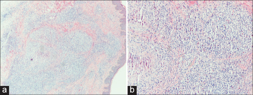
We report a 30-year-old woman who presented with a three-year history of itchy and tender lesions over the left ear, mastoid region and adjacent scalp. These were increasing in size. Cutaneous examination revealed bright red, eruptive papules and nodules ranging in size from 0.5 cm to 1 cm in diameter [Figure - 1]a and [Figure - 1]b. Examination showed no regional lymphadenopathy or any other abnormalities. The routine blood chemistry and urine analysis were within normal limits. The absolute eosinophil count and immunoglobulin E levels were normal. Histopathological examination of a nodule revealed mild epidermal hyperplasia and proliferation of small blood vessels lined by large endothelial cells. There was a dense perivascular infiltrate of lymphocytes and eosinophils [Figure - 2]a and [Figure - 2]b. The clinical and histopathological findings confirmed the diagnosis of angiolymphoid hyperplasia with eosinophilia.
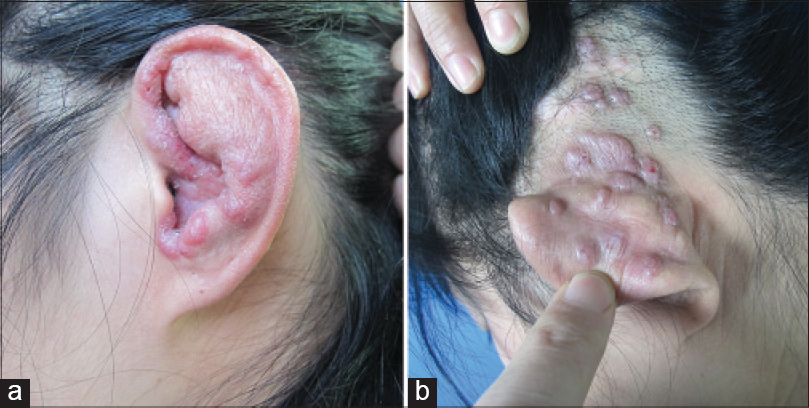
Figure 1: (a and b) Grouped plaques and nodules involving the skin of the left ear, posterior auricular area and adjacent scalp
Figure 2: (a and b) Histopathology shows prominent vascular endothelium and perivascular inflammatory infiltration, accompanied by eosinophils. (a: H and E, ×100, b: H and E, ×200)

We decided to treat the patient with surgical excision and electrocoagulation. Surgical excision was chosen for the scalp lesions because it is considered to be the most effective treatment modality.[2] However, it was difficult to excise the lesions over the ear and mastoid region. Moreover, multiple lesions are difficult to suture. Therefore, we decided to electrocoagulate those lesions using bipolar forceps [Figure - 3]a and [Figure - 3]b.
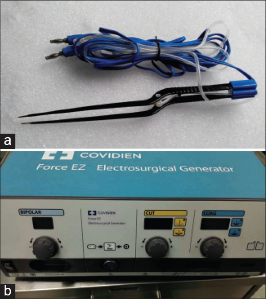
Figure 3: (a) The bipolar coagulation forceps. (b) The electrosurgical generator to which bipolar coagulation forceps was connected

Half of the lesions over the left ear and mastoid region were electrocoagulated in the first session. The second session was performed two months later to reduce the chances of necrosis in that region. There was some pain over the ear for a few days after treatment. The lesions cleared after two sessions and were no longer pruritic. Some of the lesions healed with depigmentation. There was no recurrence over a follow-up period of nine months [Figure - 4]a and [Figure - 4]b. Overall, the patient was satisfied with the outcome.
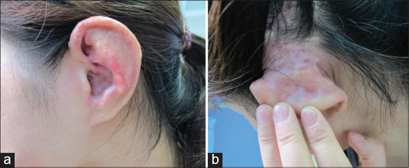
Figure 4: (a and b) Appearance of the lesions at nine months follow-up

Angiolymphoid hyperplasia with eosinophilia was first reported in 1969 by Wells and Whimster. It is a slowly progressing and self-limiting disease.[1] We should distinguish this disorder from Kimura's disease, angiosarcoma, Kaposi sarcoma and bacillary angiomatosis. Kimura's disease is characterized by vascular proliferation, lymphocytic nodules with subcutaneous germinal foci, fibrosis and edema with deep inflammation. It presents with broad subcutaneous masses, mostly in young adults of Asian descent. In contrast to Kimura's disease, angiolymphoid hyperplasia with eosinophilia may be seen in every race. The lesions are smaller and more superficial. In Kimura's disease, the histopathology shows T-cell lymphoid aggregates with well-formed B-cell germinal foci. However, in angiolymphoid hyperplasia with eosinophilia, there is nodular and diffuse T cell infiltration with small clusters of B-cells. Additionally, Kimura's disease can be differentiated from angiolymphoid hyperplasia by the presence of vascular proliferation and the endothelial cells usually being flat.

Many treatment modalities have been used in angiolymphoid hyperplasia with eosinophilia. Recently, intralesional radiofrequency ablation has been tried in a few cases. There was no recurrence after 4 to 66 months of follow-up.[4] However, electrocoagulation is easier to perform with bipolar coagulation forceps compared to intralesional radiofrequency and other treatment modalities. We were unable to find any previous reports of the use of this novel treatment modality in angiolymphoid hyperplasia with eosinophilia.

Although it may do some harm to adjacent normal tissues, it is safer because the high-frequency electrical energy produced is limited to the region between the two ends of forceps. It can be used at cosmetically important sites with multiple lesions, and is easy to perform. However, it may destroy the shape of the ear to a certain extent. It is important to control the depth of the procedure when using the coagulation forceps and to be careful in areas where important structures such as major nerves and vessels are present underneath.

Financial support and sponsorship

Nil.

Conflicts of interest

There are no conflicts of interest.

References
1.
Wells GC, Whimster IW. Subcutaneous angiolymphoid hyperplasia with eosinophilia. Br J Dermatol 1969;81:1-14.
[Google Scholar]
2.
Cheney ML, Googe P, Bhatt S, Hibberd PL. Angiolymphoid hyperplasia with eosinophilia (histiocytoid hemangioma): Evaluation of treatment options. Ann Otol Rhinol Laryngol 1993;102(4 Pt 1):303-8.
[Google Scholar]
3.
Chong WS, Thomas A, Goh CL. Kimura's disease and angiolymphoid hyperplasia with eosinophilia: Two disease entities in the same patient: Case report and review of the literature. Int J Dermatol 2006;45:139-45.
[Google Scholar]
4.
Singh S, Dayal M, Walia R, Arava S, Sharma R, Gupta S. Intralesional radiofrequency ablation for nodular angiolymphoid hyperplasia on forehead: A minimally invasive approach. Indian J Dermatol Venereol Leprol 2014;80:419-21.
[Google Scholar]

Fulltext Views
4,318

PDF downloads
1,770
Show Sections